Case submitted and written by Dr. Mazen El-Baba, with edits from Dr Jesse McLaren
As you read this, remember that the PMCardio Queen of Hearts AI ECG Model is now FDA approved. Your patient need not sit around any more with dying myocardium, and your interventionalist need not be called in for false positives. The solution is here and available.
See more here: Queen of Hearts Now FDA Approved!! What’s her take on this “Non-diagnostic” ECG?
Case
A 50-year-old male with a 40-pack year smoking history by EMS with sudden-onset midsternal chest pain followed by syncope. The pain had started approximately six hours prior to arrival, and radiated to his jaw, and was associated with shortness of breath, dizziness, diaphoresis, and a witnessed syncopal episode. He had not taken aspirin before arrival. The pain had been 10/10 at home and had improved to 6/10 by the time he arrived in the ED.
On triage assessment, he continued to have chest pain (2/10). His blood pressure was 126/85 mmHg, heart rate 67 beats/min, respiratory rate 18 breaths/min, oxygen saturation 96% on room air, and temperature 37°C. Heart sounds were normal, the lungs were clear, and radial pulses were equal.
The first ECG was recorded at triage. What do you think?
.
.
.
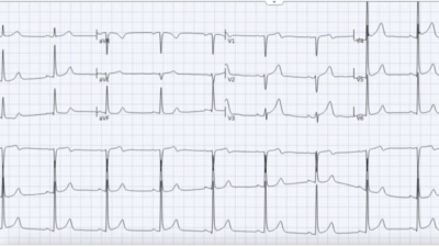
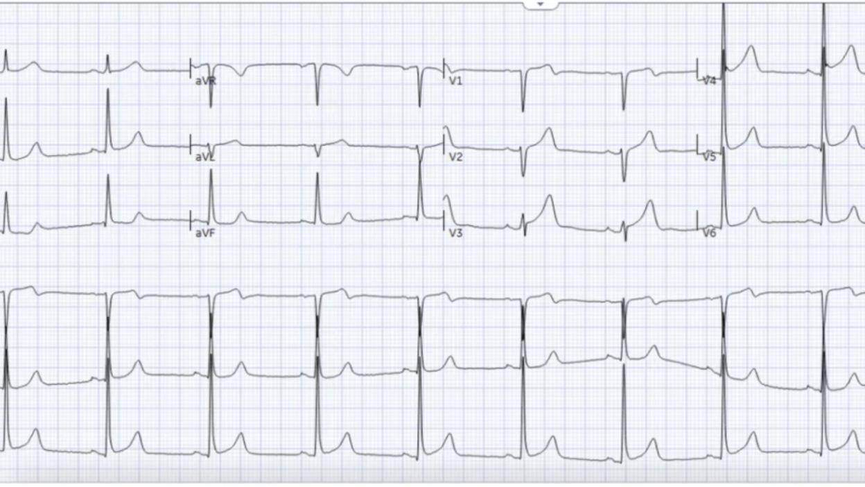
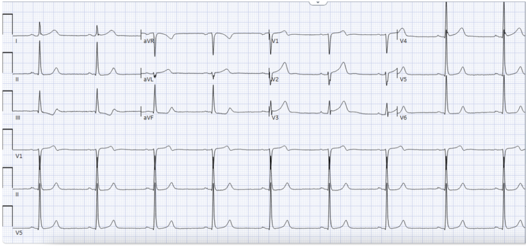
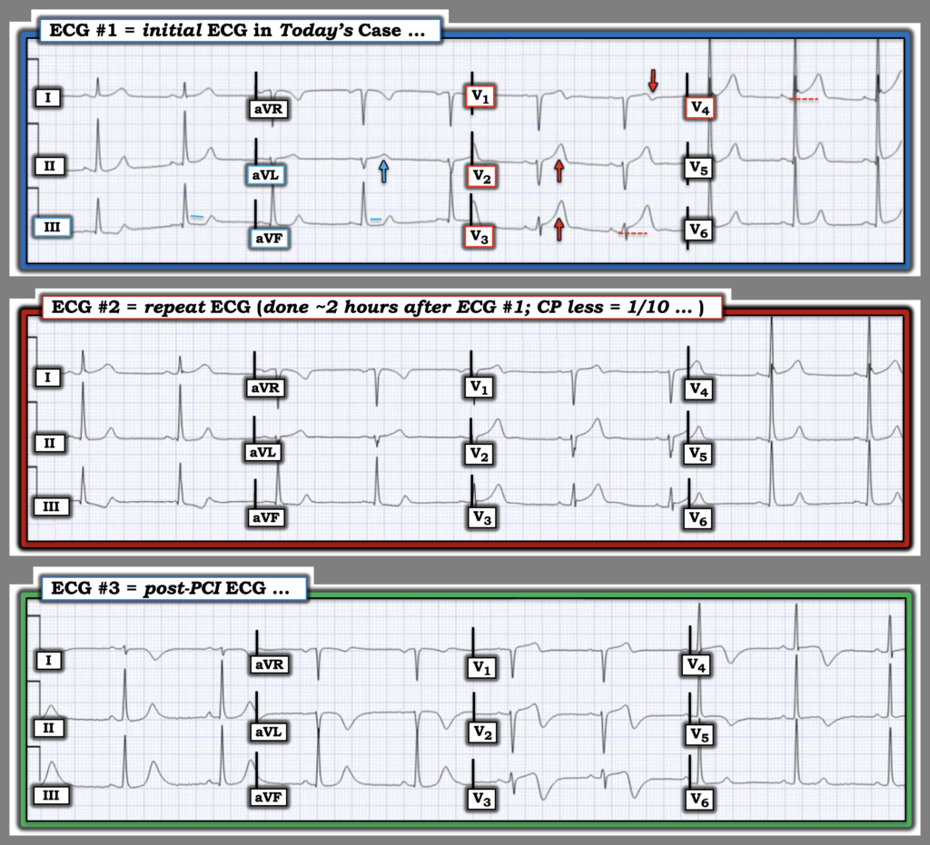
The ECG shows normal sinus rhythm, normal intervals/normal axis/R-wave progression, and tall voltages in the left sided leads that make the anterior leads more difficult to interpret. There’s subtle down/up T wave in III that could be early reciprocal change to high lateral.
Smith: I was only convinced when I saw this down-up T-wave in lead III. I think it makes the ECG diagnostic of OMI.
The ECG was signed off as STEMI negative, and the patient waited for a high-sensitivity troponin, which was 1,665 ng/L. He was then seen by the ED physician, 2 hours post-arrival, when chest pain had improved to 1/10, with a repeat ECG.
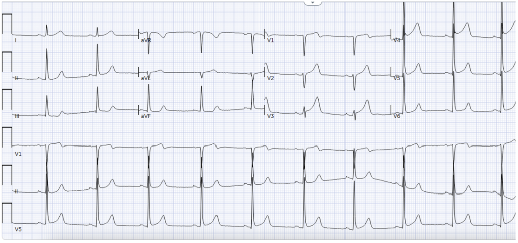
Still STEMI negative, but there’s dynamic and now more obvious down/up T wave inferiorly. Despite improvement in pain the ECG looks more ischemic, not less. Bedside echocardiography was performed, showing extensive regional dysfunction: the apex and the mid-to-distal anterior, lateral, and septal walls appeared akinetic. This was highly concerning for acute LAD-territory ischemia. Here’s the parasternal short view:
Non-STEMI or STEMI(-)OMI?
At this point, the working diagnosis should be OMI until proven otherwise, with immediate angiography. The emergency physician contacted cardiology and advocated for angiography based on:
- History: sudden substernal pain radiating to the jaw, with diaphoresis, dyspnea, and syncope; although the pain improved from 10/10 to 6/10 to eventually 1/10, it had not completely resolved
- ECG: subtle dynamic ECG changes
- Troponin: troponin 1,665ng/L
- POCUS: extensive LAD-territory regional wall-motion abnormalities.
But because serial ECGs were ‘STEMI negative’, cardiology admitted the patient as ‘Non-STEMI.’ This plan did not change when repeat high-sensitivity troponin increased to 6,557 ng/L. The troponin continued to rise, reaching 21,018 ng/L the following day.
Smith: a rising troponin is not proof of persistent OMI. It means that there was occlusion earlier and possibly continuing. Only the ECG can tell you if there is persistent occlusion. The presence of persistent chest pain is helpful (and a guideline indication for emergent reperfusion!), but is not always present with persistent occlusion — the ECG is more reliable and the patient should have been taken to the lab on the first ECG.
This marked rise prompted coronary angiography at approximately 19 hours after his initial presentation. Angiography showed a 99% proximal LAD occlusion with TIMI 1 flow, treated with PCI.
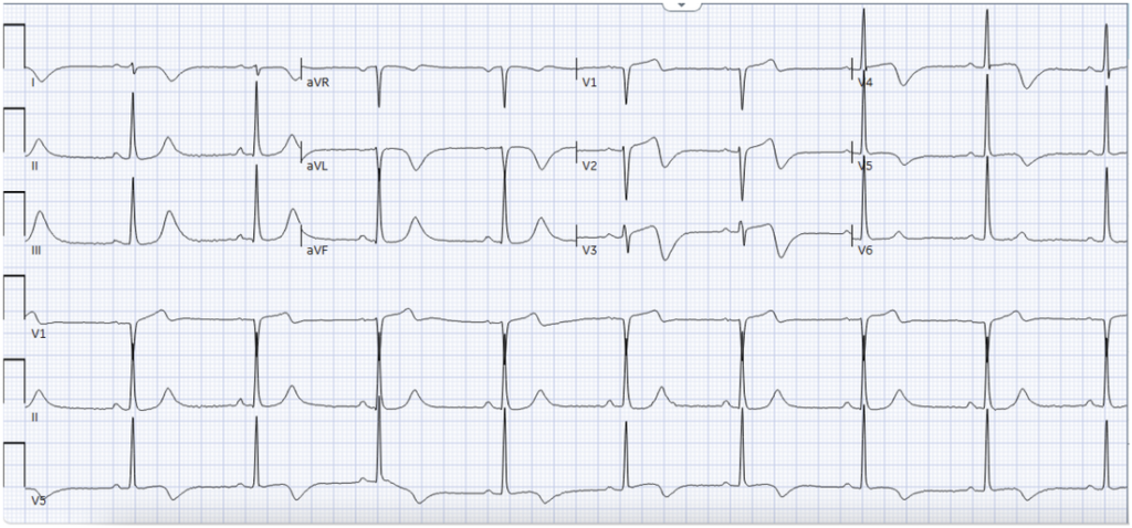
An ECG obtained after PCI showed reperfusion T wave inversion in the distribution of the proximal LAD:
Formal echocardiography after angiography showed an LVEF of 35–40%, with an akinetic apex and akinesis of the mid-to-distal anterior, lateral, and septal walls:
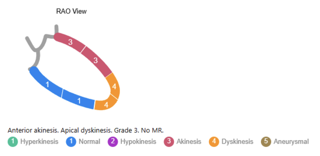
STEMI vs OMI paradigms
According to the STEMI paradigm this was a ‘Non-STEMI’. Guidelines recommend an inpatient invasive strategy for intermediate- or high-risk NSTE-ACS, with angiography within 24 hours considered appropriate for many high-risk patients. Angiography at approximately 19 hours therefore fell within a conventional NSTEMI pathway (though if pain is refractory, there should be immediate angiography, based on NSTEMI guidelines that are rarely followed). This means this case will not be flagged for quality improvement. In the STEMI paradigm, the discharge diagnosis changes for ‘false positive STEMI’ but not false negatives: the ‘NSTEMI’ diagnosis remains even in hindsight, so occlusions are missed both prospectively and retrospectively.[1]
But this was a STEMI-negative OMI. This patient had persistent ischemic symptoms and objective evidence of regional myocardial dysfunction while still in the ED. Waiting for STEMI criteria or a larger troponin rise meant waiting for more myocardium to infarct.
In retrospect, earlier angiography would clearly have been preferable for a patient with TIMI-1 proximal LAD occlusion and EF reduced to 35%. And this STEMI(-)OMI could have had rapid reperfusion based on datapoints that go beyond STEMI criteria: the patient had a highly concerning history, residual ischemic pain, a dynamic ECG abnormality, a troponin rise from 1,665ng/L, and extensive LAD-territory regional dysfunction on POCUS. Together, these findings created a compelling case for OMI and for immediate same-night angiography rather than waiting for the troponin to continually rise.
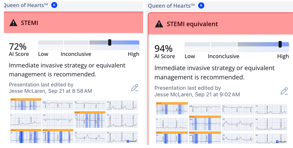
AI-trained on OMI can help the reperfusion decision by identifying subtle but important ECG changes. Here’s the Queen of Hearts interpretation of the two ECGs, with different interpretations for V2, but identifying the inferior down-up T waves reciprocal to subtle high lateral hyperacute T waves, with greater confidence on the second ECG:
Take-home points
- OMI is a clinical diagnosis supported by multiple data points. History establishes the pretest probability; ECG, serial ECGs, troponin, and cardiac imaging update it.
- Subtle ECG findings matter, especially when they are dynamic, and reciprocal change may be earlier and more obvious .
- Improving pain is not the same as resolved pain. Even 1/10 pain = ongoing pain, and ACS with refractory pain requires immediate angiography
- Troponin diagnosis myocardial injury, not coronary patency. Do not wait for a large rise, or use the label NSTEMI, to decide whether threatened myocardium needs immediate reperfusion.
- POCUS can change the probability. A normal POCUS cannot rule out OMI, but in a high-risk patient with subtle ECG changes, a new regional wall-motion abnormality strongly supports OMI.
- Guideline-concordant timing based on STEMI paradigm leads to reperfusion delays for STEMI(-)OMI, which can be prevented by attention to clinical, ECG and POCUS features of OMI
- Ischemic pain + dynamic ECG change + regional wall-motion abnormality = OMI until proven otherwise.
Reference
- McLaren, El-Baba, Sivashanmugathas, Meyers, Smith, Chartier. Missing occlusions: quality gaps for ED patients with occlusion MI. AJEM 2023
Smith’s comments:
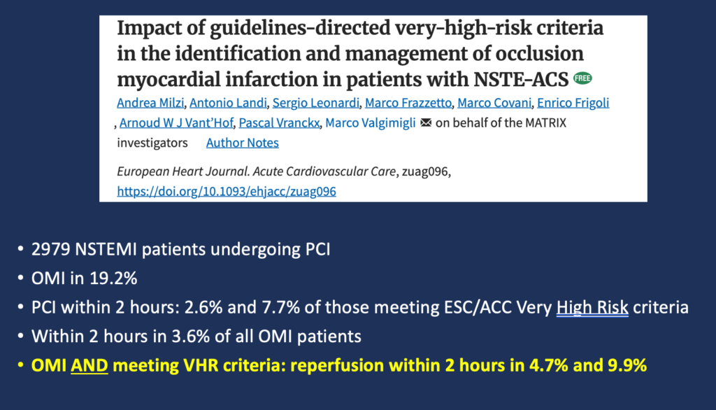
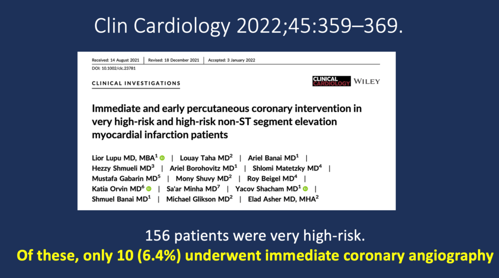
The NSTEMI guidelines do not work. In Lupu et al, 6% of patients who met “high risk criteria” had the guidelines followed. In a new study by Milzi et al. fewer than 10% of OMI that met high risk criteria had guideline acceptable care. “High Risk” NSTEMI, with persistent myocardial loss, pulmonary edema, electrical instability, and shock/hypotension is actually “Complications of Undiagnosed OMI”. As is the case throughout the world, NSTEMI “high risk” guidelines are not followed and, even if they were, the intervention would be too slow to save much myocardium, since the high risk guidelines usually depend on troponin elevation (damage done) with or without complications. That is why we must diagnosis OMI on the first ECG, activate the cath lab, and get the artery open to save myocardium, save lives, and prevent heart failure. This can only be reliable done using the PMCardio Queen of Hearts AI ECG Model.
Remember: NSTEMI is a worthless diagnosis.
References:
Lupu L, Taha L, Banai A, et al. Immediate and early percutaneous coronary intervention in very high-risk and high-risk non-ST segment elevation myocardial infarction patients. Clin Cardiol [Internet] 2022;Available from: https://onlinelibrary.wiley.com/doi/10.1002/clc.23781Milzi A, Landi A, Leonardi S, et al. Impact of guidelines-directed very-high-risk criteria in the identification and management of occlusion myocardial infarction in patients with NSTE-ACS. Eur Heart J Acute Cardiovasc Care [Internet] 2026;(zuag096). Available from: http://dx.doi.org/10.1093/ehjacc/zuag096/8764093
= = =
==================================
MY Comment, by KEN GRAUER, MD (9/24/2026):
I found it disturbing to review today’s case. For example:
- The patient is a 50-year old man with the strong risk factor of heavy longterm smoking — who describes the sudden onset of 10/10 CP (Chest Pain) that prompts him to go to the ED.
- The initial ECG is “signed off as STEMI-negative”.
- The patient then “waited for a high-sensitivity Troponin”.
- The patient’s CP had decreased from 10/10 at its onset — to 6/10 by the time he arrived in the ED (ie, at the time ECG #1 was recorded) — and finally to 1/10 during the time he was “waiting for a hs-Troponin” to come back.
- The patient was “then” seen by the ED Physician (ie, The patient was apparently only seen 2 hours after his arrival in the ED — which was only after the 1st Troponin came back significantly elevated at 1,665 ng/l — which apparently, only then prompted the recording of a 2nd ECG).
The ED physician appropriately obtained bedside Echo (which showed extensive regional LV dysfunction). The ED physician appropriately recognized subtle “dynamic“ ECG changes on ECG #2 — and appropriately contacted cardiology, advocating for prompt cath based on all of the above. But cardiology “stuck” to the “STEMI-negative” label — such that cardiac cath showing acute proximal LAD occlusion was not performed until the next day (= 19 hours after the patient’s arrival in the ED).
- Drs. Mazen El-Baba and McLaren highlight the reasons why all of the above parts of today’s case are disturbing.
= = =
The ECGs in Figure-1:
“STEMI-negative” is the wrong description for today’s initial ECG. That’s because the history ( = sudden onset of 10/10 CP in a high-risk patient — but with significant reduction in severity of symptoms by the time ECG #1 is recorded) tells us that we may not see the ST elevation required to satisfy STEMI criteria. But regardless of whether or not the initial ECG satisfies STEMI criteria — there is clear indication in ECG #1 of an acute OMI until proven otherwise:
- Our “eye” should be immediately captured by the disproportionately increased hyperacute T wave in lead V3 (that dwarfs the tiny QRS complex in this lead). There is also a small-but-definite amount of inappropriate ST elevation in this lead.
- Neighboring lead V4 shows a bit more ST elevation (which we know is abnormal — because there is no more than trivial ST elevation in the subsequent more lateral chest leads V5 and V6).
- Neighboring lead V2 also shows a hyperacute (disproportionately increased) T wave considering the small QRS complex in this lead.
- The subtle-but-steep decline of the small but positive T wave in lead V1 into terminal negativity (RED arrow in this lead) is not a normal finding. In the clinical context of reduced CP ( = 6/10 at the time this tracing is recorded) — this finding suggests at least some degree of spontaneous reperfusion as the reason for the patient’s reduced CP.
- Limb lead findings in ECG #1 are also subtle — but they are consistent with the diagnosis of acute OMI until proven otherwise (ie, There is a disproportionately increased T wave in lead aVL — and ST segment straightening in inferior leads III and aVF).
As per Drs. Mazen El-Baba and McLaren — ECG #2 shows subtle-but-real “dynamic” ST-T wave changes in the form of increased acuity of the angulation of ST depression in leads III and aVF (that would be EASY to miss if one did not interpret ECG #2 by placing it right next to ECG #1).
- Confirmation that the findings I describe above in ECG #1 are real — is forthcoming from inspection of ECG #3 — that shows dramatic reperfusion T waves in leads V1,V2,V3,V4 — as well as in limb leads aVL, III and aVF (which are all of the leads showing signs of what was to come in the initial ECG).
Bottom Line: Review of the events in today’s case is disturbing …
= = =
Figure-1: Comparison between the 3 ECGs in today’s case.
= = =
= = =