Today’s case is a repost from October 26, 2010. I have updated the post — and Ken Grauer has added a new Comment below.
This 54 year old patient with a history of kidney transplant and poor transplant function — had been vomiting all day, when at 10 PM he developed severe substernal crushing chest pain.
- He presented to the Emergency Department with a blood pressure of 111/66 and a pulse of 117.
= = =
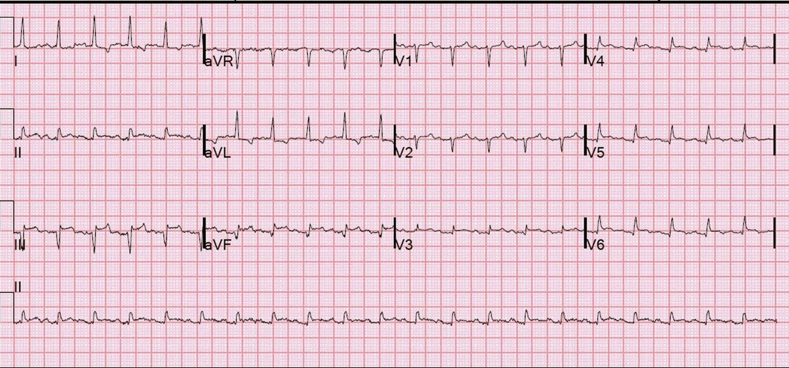
He had this ECG recorded.
- He was rushed by residents into our critical care room with a diagnosis of STEMI (STEMI-OMI), and they handed me this ECG:
= = =
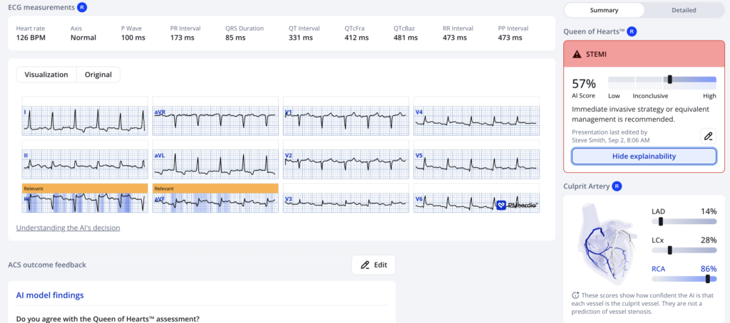
There is sinus tachycardia with ST elevation in II, III, and aVF, as well as in leads V3-V6. There is reciprocal ST depression in I and aVL. At first glance, it seems the patient is having a STEMI.
In fact, the cath lab had already been activated before the patient was in my care.
Remember, we do not evaluate and treat ECGs; we evaluate and treat patients. Even if this ECG is the first thing one sees (as it was for me) — one should stop and think: “This is an unusual Occlusion MI.” Why?
ACS and OMI generally do not cause tachycardia unless there is cardiogenic shock. Are the lungs clear? Is the patient cool and pale? Then ACS (STEMI) might be primary; this might be cardiogenic shock.
Most importantly, what does the bedside echo show for cardiac function?
More often, tachycardia with ST segment abnormalities (elevation or depression) — is due to an underlying illness (PE, sepsis, hemorrhage, dehydration, hypoxia, respiratory failure, etc.). One must clearly rule out these processes before jumping on the ACS diagnosis.
Furthermore, notice the well-formed Q-waves in the inferior leads. These must raise suspicion of old MI with persistent ST elevation.
One very useful adjunct is ultrasound:
- Echo of his heart can distinguish aneurysm from acute MI by the presence of diastolic dyskinesis, but it cannot distinguish demand ischemia from ACS.
- In today’s case — bedside echo did not reveal any visible wall motion abnormality. Instead,there was hyperdynamic function, which is not consistent with cardiogenic shock, but rather with sepsis or volume deficit. Wall motion can be very tricky to perceive, especially in tachycardia.
Large volume fluid resuscitation was undertaken.
- The K returned at 6.9 mEq/L. The HCO3 was 8. Cr was 13.4. Even after 3 liters of fluid, his CVP was very low.
Troponins (4th generation, not hs) peaked at 0.275 ng/ml, consistent with myocardial injury, but not necessarily myocardial infarction. And if there is myocardial infarction it is 1) quite small and 2) likely type 2 MI from supply demand mismatch, NOT from ACS.
(Type 2 MI is called “Secondary MI” in the 5th Universal definition of MI.)
As the cath lab had been activated, the patient did undergo an angiogram which showed no acute coronary lesions.
The patient was suffering from severe dehydration, possibly with sepsis.
The Queen of Hearts is not a clinician, and evaluates the ECG only, not the patient. Thus, one cannot blame her for calling this OMI with low confidence (57% is low confidence).
After stabilization, previous ECGs and an old echocardiogram were found:
- The prior ECGs demonstrated an old inferior MI, with persistent ST elevation (LV aneurysm morphology).
- The previous Echo showed diastolic dyskinesis.
= = =
LV aneurysm morphology on the ECG is also called “Persistent ST Elevation after Previous Old MI”. It seems to be far more common in anterior MI, where it presents with 1) QS-waves (Q-wave without any subsequent R-wave) and 2) mild ST elevation and either mildly upright or slightly inverted T-waves. But inferior aneurysm is much more difficult to recognize because it often has a QR-wave, as in this case.
See here for more information from this blog on LV aneurysm
==================================
MY Comment, by KEN GRAUER, MD (8/11/2026):
Today’s case is a repost of a “short-and-sweet” case from 16 years ago. That said, this post is full of “truisms“ with PEARLS that you won’t find in textbooks (with these PEARLS being equally applicable today as they were in October 26, 2010 when this case was first published). The PEARLS from today’s repost case include the following:
- We do not evaluate and “treat” ECGs. Instead — we evaluate and treat patients (Each patient that you see presents with their own unique “story” — and each patient has an initial ECG recorded on them; We then interpret that ECG in context with that patient’s unique story).
- ACS and acute MI generally do not cause tachycardia (unless there is cardiogenic shock). So if your patient with new CP (Chest Pain) presents with tachycardia but no sign of shock — then look for some cause other than ACS/MI.
- Not all patients with hyperkalemia show obvious signs of hyperkalemia. There are 2 potential reasons for this: i) Reason #1 = Just because … (ie, Because we don’t know why the sky is blue — and because we also don’t always know why some patients with hyperkalemia do not have obvious signs of this on their ECG); — and, ii) Reason #2 = The ECG shows the net effect of hyperkalemia which is superimposed on what the patient’s baseline ECG looked like. This “net effect” will also depend on whatever else might be going on (ie, If the patient’s baseline ECG shows a lot of ST depression — then this may attenuate, or even cancel out the peaked T waves of new hyperkalemia).
- We have no idea as to what a hyperkalemic patient’s “true” acute ECG will look like (until we repeat the ECG after hyperkalemia has been corrected).
- Sometimes the “peaked” T waves of hyperkalemia are subtle.
- Instead of peaked and pointed upright T waves (that we are all well accustomed to seeing) — Patients with hyperkalemia sometimes manifest inverted T waves that may be peaked and pointed.
- Not all ST elevation is from acute MI (ie, ST elevation may be the result of previous infarction, now with development of LV aneurysm — as was suspected in today’s case). Other factors may contribute (ie, ST elevation may look worse at faster heart rates).
- Finding a previous ECG may clarify (and even confirm) the diagnosis that you suspect. Once you verify that a previous ECG is a true “baseline” (ie, that this previous tracing was not taken at a time when the patient was having an acute event) — Comparing this baseline tracing with the new initial ECG may prove invaluable for distinguishing between what is “new” vs “old” vs “new superimposed on old.
- Bedside Echo is an invaluable tool for ruling in or out a series of important cardiopulmonary conditions — and it does so in minimal time! (ie, In today’s case — the hyperdynamic function and lack of any wall motion abnormality confirmed the need to look for a diagnosis other than ACS/acute MI).
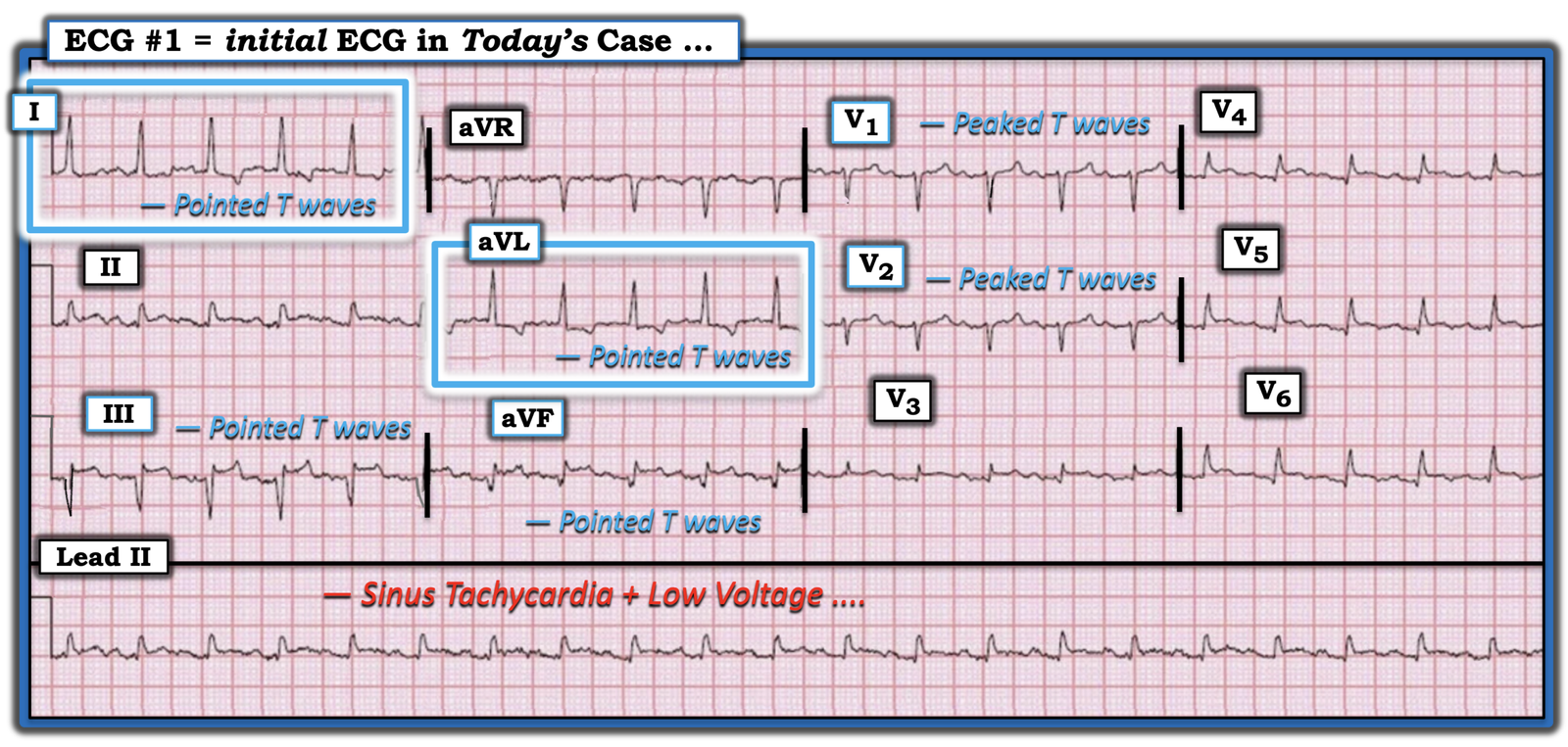
- There is surprisingly low voltage in many of the leads in today’s tracing! (See Figure-1 below — in which I have reproduced and labeled today’s ECG).
- Although low voltage may be a benign, incidental finding — there is a differential diagnosis for Low Voltage that we need to always consider, especially in a patient who presents with new CP, as in today’s case (CLICK HERE — for review of the important causes of low voltage).
- In addition to low voltage — there is also tachycardia in today’s case. The combination of low voltage + tachycardia always requires us to find out the reason why!
= = =
Today’s ECG …
I fully acknowledge that on seeing today’s ECG — I initially considered an acute OMI because of the ST elevation in the inferolateral leads.
- And then I considered the history … — for which learning about this patient’s longterm renal issues suggested the possibility of acute renal failure with associated electrolyte imbalance.
- And then I considered the above PEARLs:
- Although T waves that are both tall and pointed (or at least peaked) are not seen in today’s tracing — Not all patients with hyperkalemia show obvious signs of hyperkalemia on their ECG.
- None of the T waves in Figure-1 are tall. That said — the T waves in lead III are pointed — and the T waves in leads aVF,V1,V2 are at least peaked.
- But what most “caught my eye” — were the pointed negative T waves in leads I and aVL (within the BLUE rectangles). This is not the characteristic appearance for T waves serving as reciprocal change to an acute MI.
- Bottom Line: I immediately knew from the history and from Figure-1 that optimal management of today’s patient would require finding out as soon as possible what the serum K+ level was. Profound volume depletion soon became obvious — and study results ruled out an acute cardiac event without the need for cardiac catheterization.
= = =
Figure-1: I’ve reproduced and labeled today’s ECG.
= = =
= = =