By Matthew McArthur and Jesse McLaren
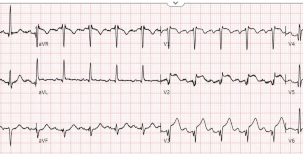
4 patients presented with ST elevation, active infection with or without fever, and elevated troponin. Which had true positive STEMI and which had myocarditis?
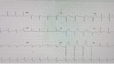
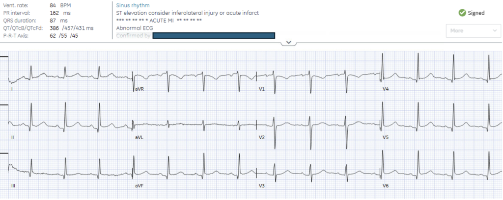
Patient 1: 70 year-old on chemotherapy, presenting with fever, nausea/vomiting, and hs-trop I of 7,000ng/L
–
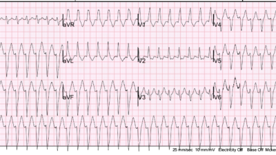
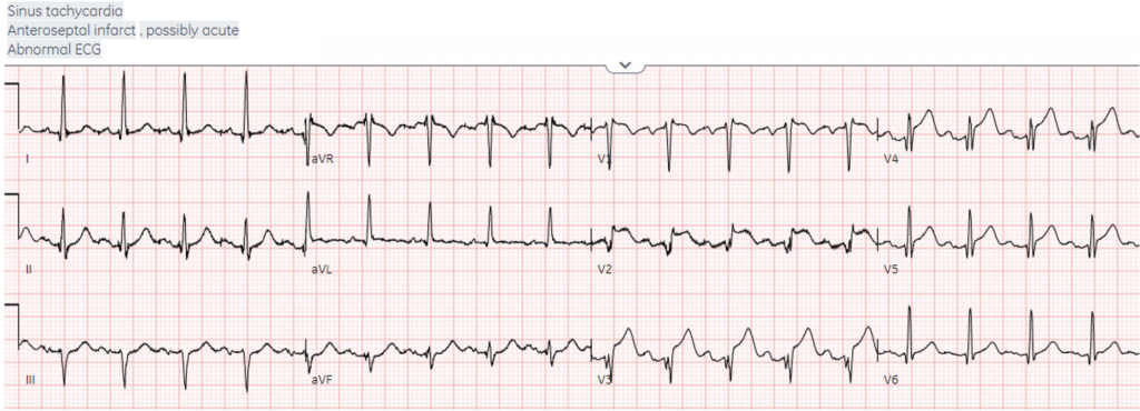
Patient 2: 55 year old recent cough and chills, presenting with 6 hours of chest pressure
–
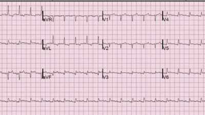
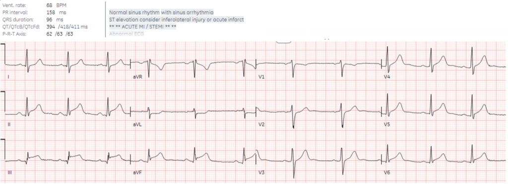
Patient 3: 25 year old, recent strep now, presenting with acute chest pain
–
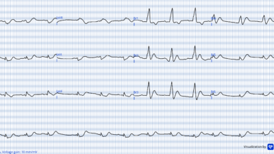
Patient 4: Healthy 30 year old, recent gastroenteritis-like illness, with acute chest pain which was positional and pleuritic. Computer interpretation read *ACUTE MI/STEMI*
STEMI vs myocarditis
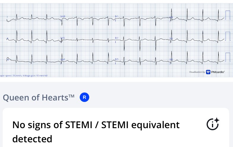
STEMI criteria has high rates of false positive activation, ie code STEMI without culprit lesion. These can be stratified by preventable false positive (eg troponin negative, where the ECG has identifiable non-ACS cause of ST elevation, eg early repolarization, pericarditis, LVH, LBBB without Modified Sgarbossa Criteria), vs appropriate false positive where the diagnosis requires angiography to exclude acute coronary occlusion (eg troponin positive with ST elevation from takotsubo, SCAD, vasospasm). Queen of Hearts (QoH) can reduce preventable false positives – including all the cases of pericarditis in this study.
Myocarditis is more challenging as troponin is positive, and there can be a range of ECG changes – from non-specific to diffuse ST elevation like pericarditis, to regional/reciprocal ST changes that mimic true positive STEMI. But the latter is an angiographic diagnosis of exclusion – including in young patients (who can get an acute coronary occlusion or SCAD), those without risk factors (until their first event), and those with viral prodromes (which are prothombotic). After an initial workup including ECG, troponin and echo, the 2024 ACC expert consensus on evaluation of myocarditis advises that “Obstructive coronary artery disease should be excluded by coronary angiography (invasive or via CT) as clinically appropriate,” followed by diagnosis by cardiac MR.
A new study found QoH can identify inflammatory myopericardial syndroms as non-OMI with high specificity. Discordant cases which were positive by STEMI criteria but had no signs of OMI detected by QoH predominantly showed diffuse non-territorial inflammatory patterns. Specificity was higher for pericarditis than myocarditis, with more QoH false positives for myocarditis than pericarditis – which is appropriate. In other words, QoH can help stratify inflammatory myopericardial syndromes into preventable false positive STEMI without signs of OMI (who may still require exclusion of obstructive coronary disease but not necessarily by emergent angiography), vs appropriate false positive STEMI with ECG that mimics OMI and remains an angiographic diagnosis of exclusion.
Back the cases
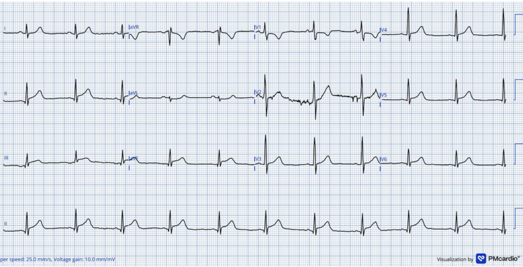
Patient 1: 70 year-old on chemotherapy, presenting with fever, nausea/vomiting, and hs-trop I of 7,000ng/L
There’s mild infero-lateral STE, II>III, with no reciprocal STD in aVL (which makes inferior OMI very unlikely) and reciprocal change in aVR only. Because of STEMI criteria + troponin elevation the cath lab was activated, but QOH sees no sign of OMI. Coronaries were normal, peak troponin was 9,000. Echo showed hypokinesis of inferolateral walls, and cardiac MR showed pericardial edema and effusion. Diagnosed with perimyocarditis. Discharge ECG showed normalization:
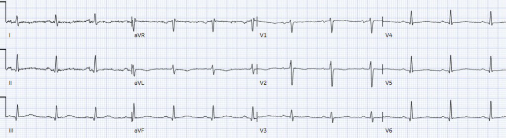
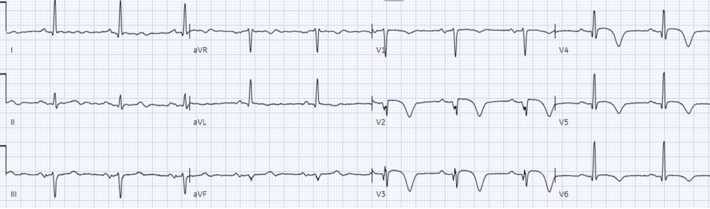
Patient 2: 55 year old recent fever and chills, presenting with 6 hours of chest pressure
There’s sinus tachycardia, Q waves anteriorly with STE and hyperacute T waves, without lateral or inferior changes – corresponding to mid LAD occlusion, identified as OMI by QoH. Cath lab activated: 100% mid LAD occlusion. First hs-trop I was 1,000ng/L and peak 90,000, with echo showing anterior hypokinesis. Also tested positive for COVID. Discharge ECG showed ongoing anterior Q waves, and reperfusion T wave inversion:
Smith: the first ECG in this case does not look at all like myocarditis to me and the reason for that is that the T-waves are hyperacute. Myocarditis has ST Elevation, but not hyperacute T-waves. Some OMI also does not have hyperacute T-waves, and these are the cases that could be either OMI or myocarditis. But when there are hyperacute T-waves, it is OMI until proven otherwise!
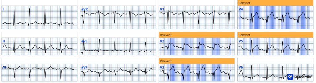
Patient 3: 25 year old, recent strep now, presenting with acute chest pain
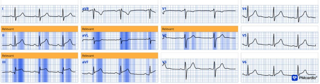
There’s inferior STE with reciprocal STD-aVL, and mild anterior STD – concerning for infero-posterior STEMI(+)OMI – identified by QOH as OMI. Cath lab was (appropriately) activated, which excluded acute coronary occlusion. First hs-trop I was 500 and peak 7,000 ng/L. Echo was normal, with no regional wall motion abnormalities or pericardial effusion. Cardiac MR showed inferolateral subepicardial enhancement with adjacent small pericardial effusion. Diagnosed with myocarditis. Discharge ECG showed normalization:
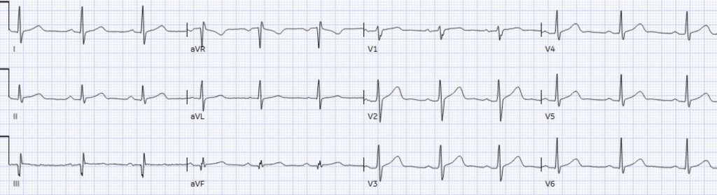
Patient 4: Healthy 30 year old, recent gastroenteritis-like illness, with acute chest pain which was positional and pleuritic. Computer interpretation read *ACUTE MI/STEMI*
There are contiguous ST elevations in the inferior leads and lateral precordial leads. There is reciprocal ST depression in aVL. Of course, this is concerning for inferolateral STEMI. QOH called STEMI with 87% AI score.
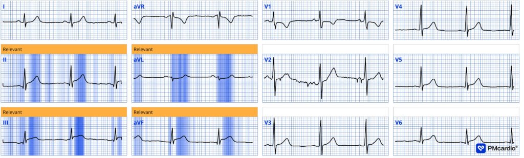
Because of age and viral prodrome, the patient received ASA in ED triage 80 minutes prior to assessment and their chest pain had since improved from 8/10 to 1/10 in severity. Repeat ECG:
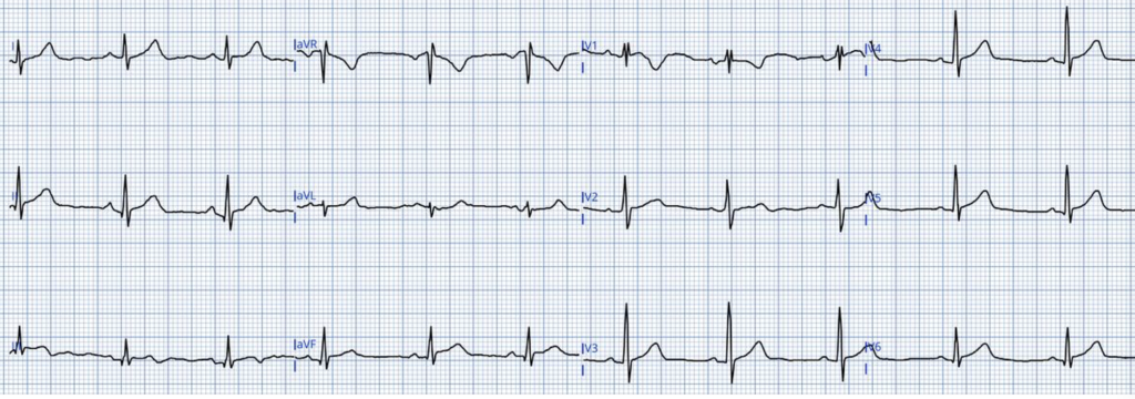
There are still inferior elevations with reciprocal depression in aVL, but now there is terminal T wave inversion in III which can be seen in reperfused OMI – QOH called this reperfused OMI with an AI score of 51%. Bedside ultrasound showed a normal LVEF, no pericardial effusion, no obvious RWMA, grossly normal RV with no RV strain. High sensitivity troponin I was 4000ng/L (normal <20).
I felt the most likely diagnosis was viral myocarditis but acute (?reperfused from ASA) inferior OMI was not excluded, particularly based on the ECG findings — so I paged the on-call STEMI physician. After reviewing the case and ECGs, they felt the case did not merit emergent cath lab activation and advised consulting internal medicine for medical management of myocarditis vs “NSTEMI”. The patient was then assessed by the internist on call who felt that the case was most consistent with myopericarditis. The patient was admitted and treated with NSAIDs and colchicine. Troponin peaked at 11,000 the following day, and CRP was initially mildly elevated at 19 (normal <5) and trended down to 12 in hospital. Inpatient echo confirmed normal biventricular function with no pericardial effusion and no wall motion abnormalities. The patient was discharged after a few days with plan for repeat outpatient echo in a few weeks.
However, there was no angiogram or coronary CT to exclude obstructive coronary disease, and no cardiac MRI to confirm myocarditis. This is a risky decision to assume a viral entity treated with anti-inflammatories, rather than excluding thrombotic occlusion that can spontaneously reperfuse but just as easily spontaneously reocclude.
Smith: the evolution, with rapid resolution of pain and terminal T-wave inversion is virtually diagnostic in this last case, and not at all typical of myocarditis. It has a very high probability of being OMI with reperfusion. I suspect that the appropriate diligence was not done because the patient is young.
OMI is not rare in young patients, and not even in young women.
Look at this case especially: A teenager with chest pain, a troponin below the limit of detection, and “benign early repolarization”
Take home
- Young age, lack of risk factors, or viral prodrome do not exclude acute coronary occlusion – see these cases of a teenager with LAD occlusion and a 30 year old with transient OMI
- (myo)Pericarditis which is ‘STEMI positive’ but without signs of OMI is a preventable false positive STEMI
- (peri)Myocarditis which is ‘STEMI positive’ and mimics OMI remains an angiographic diagnosis of exclusion
= = =
==================================
MY Comment, by KEN GRAUER, MD (8/17/2026):
In the early days of my training, and then as a young faculty member — the entity of acute pericarditis “captivated” me — with its anticipated evolution over 4 specific stages (= Diffuse ST elevation — Pseudonormalization — Diffuse T wave inversion — and Return to a normal ECG). I eventually learned the realities:
- Pure acute pericarditis is rare in clinical practice. Far more common as a presentation for emergency care is acute ischemia/OMI — so much so, that Dr. Smith’s adage, “You diagnose acute pericarditis at your Peril” — echoes through my mind as a “mantra” each time I contemplate considering the diagnosis of acute pericarditis. And then when I look further — I almost always come to the realization that the patient does not have pericarditis. To be sure — We have published cases in Dr. Smith’s Blog of true pericarditis. But these cases of “pure” pericarditis are rare.
- Instead, much (most) of the time — rather than “pure” pericarditis or “pure” myocarditis — the patient has myopericarditis (ie, some combination involving both pericardium and myocardium).
- Myopericarditis is more common than “pure” pericarditis — although among patients presenting for emergency care, myopericarditis is still much less common than acute coronary syndromes.
- Despite the above truisms — acute pericarditis still tends to be overdiagnosed. This is unfortunate — because the morbidity and mortality of missing a case of “pure” pericarditis is relatively minor compared to the potential life-threatening mistake of misclassifying an acute OMI as “pericarditis”.
- To emphasize — the morbidity of missing acute myopericarditis is not negligible; But it still is less than missing an acute OMI.
= = =
About Today’s Post …
Dr. McClaren puts the above concepts to the test — as he challenges us to decide between perimyocarditis vs acute OMI from 4 clinical vignettes and the initial ECGs that accompany them. As reinforcement — I’ll restate the “Take-Home” Points that I got from Dr. McClaren’s excellent presentation:
- We need to accept that among patients who present for emergency care with chest pain — that the initial ECG by itself may not allow us to rule out an acute cardiac event. That’s because acute myocarditis is regularly associated with elevated Troponins and ECG patterns that may mimic acute OMI. Cardiac cath may be needed for differentiation — and it is perfectly appropriate to activate the cath lab in such cases.
- Among those cases in which findings on the initial ECG do allow us to minimize the likelihood of an acute cardiac event — perhaps the best clue is when you see an ECG similar to that of Patient #1 — in which the complete lack of reciprocal ST depression in lead aVL all but negated the likelihood of acute inferior OMI (ie, When the ECG is not at all suggestive of the expected anatomic appearance for acute occlusion of a given “culprit” artery).
- Additional testing may still be needed for confirmation or to rule out an acute cardiac event (ie, Serial ECGs; serial Troponins; Echo at the bedside) — but a false positive cath lab activation can often be avoided by not immediately activating the cath lab when the initial ECG does not suggest occlusion of a specific “culprit” artery.
- If doubt remains in the mind of the provider — QoH has been shown to be highly accurate to assist for distinguishing OMIs from non-OMIs (ie, The provider can gain confidence in their decision not to immediately activate the cath lab if QoH also says, “No OMI” ).
- The History is very important! Although the prevalence of pericarditis and myopericarditis is low among all comers who present with chest pain for emergency care — younger age and eliciting a history of fever, viral symptomatolgy, chest trauma, use of certain medications (ie, medications such as chemo- or immunotherapy, or lupus-inducing drugs with known association predisposing to myopericarditis) — should at least enhance our receptivity to myopericarditis as a possibility (as did the younger age and viral prodrome that lessened the likelihood of acute OMI for Patient #4).
= = =
Bottom Line:
- We want to do our best for arriving at a definite diagnosis — always mindful (as per Dr. McClaren) — that “younger age, lack of risk factors and even viral prodrome do not exclude acute coronary occlusion”.
- As a result — ruling out acute coronary occlusion will often entail serial ECGs, serial Troponins, and bedside Echo.
- Sometimes — Coronary CT, cardiac MRI and even cardiac catheterization may be needed before sending the patient home.
= = =
= = =