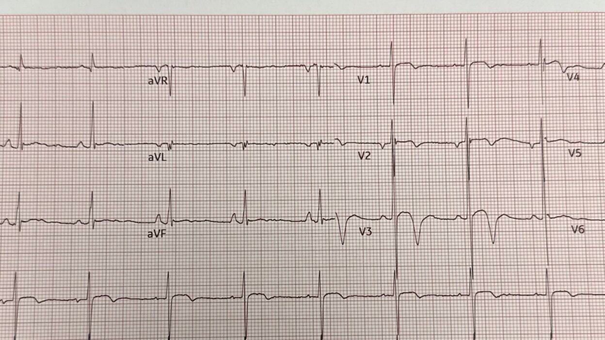
This was sent by a fine former resident, BG Tenbrink.
A 20-something male had syncope while playing basketball. There was no chest discomfort, no SOB, no symptoms in the ED.
A thorough history is available:
He was in his normal state of health. He was playing basketball and had just started the game when the ball went out of bounds. He was walking over towards the side when he began to feel lightheaded, and “the world was going dark.” He then sat down for a few moments. Moments later play resumed, and he tried to run down the court further. He was witnessed to run a few feet forward and then run into a fence and fall and hit his head. He does not remember this. He states he was down for about 2 to 3 minutes. There was no seizure activity. After this, he remembers awakening and knowing where he was, and who the people were around him. He never had any chest pain or palpitations. He says he did not eat anything before the game, but that is not unusual. He has no acute or chronic illnesses. He takes no medications. He has no drug or alcohol use.
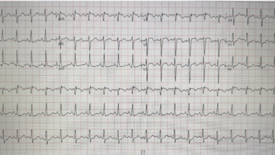
Here is his ECG, which was texted to me:
What do you think?
I immediately recognized this as a normal variant. This is classic pattern which I call “benign T-wave inversion.” It is a mimic of Wellens’ pattern. It is primarily seen in young, thin, athletic, African American men. See more info below.
Indeed, this patient was African American.
My response was that this is a normal variant in a young African American and that the ECG shows no evidence of ischemia. I wrote that hypertrophic cardiomyopathy is possible, and that an echo is indicated (even a good POC bedside echo would be adequate)
I have never seen the Queen of Hearts call this OMI. In this case, she did.
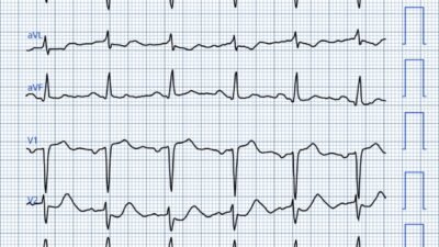
This is what she says if you answer that the symptoms are worrisome for ACS:
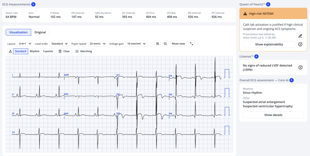
However, in this case, the symptoms are not at all typical of ACS. Syncope without other symptoms is very low risk for ACS, though there are times when syncope alone is the only symptom. In such cases, the ECG must be very specific for OMI when the symptoms give a low pretest probability.
See this article here (sorry, paywall): McLaren JTT, Smith SW. A Bayesian approach to acute coronary occlusion. J Electrocardiol 2023;81:300–2.
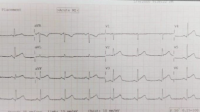
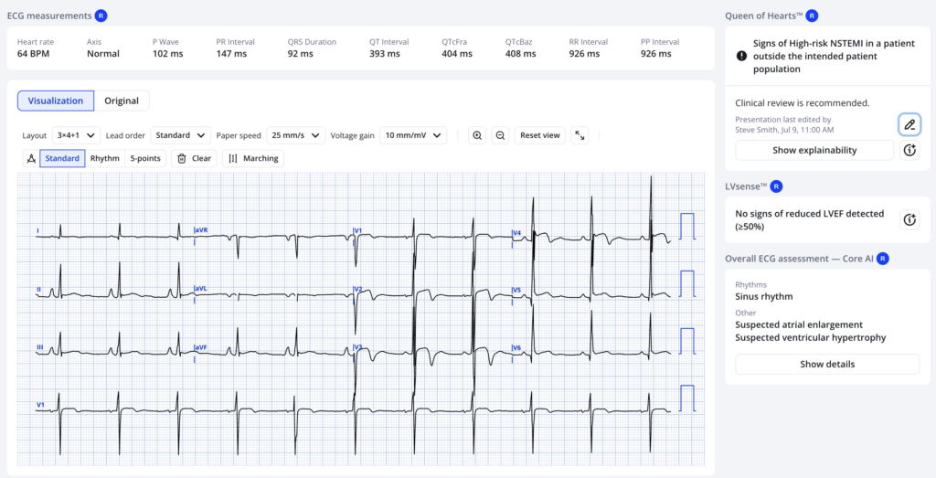
If you answer that the symptoms are not worrisome for ACS, the PMCardio Queen of Hearts gives this answer:
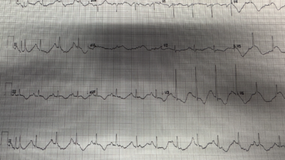
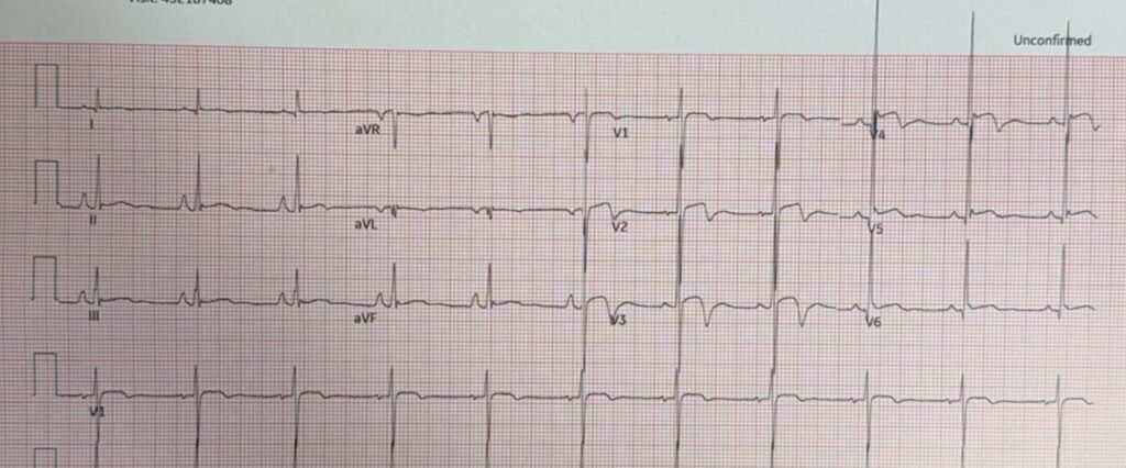
There was an old ECG available from one year prior:
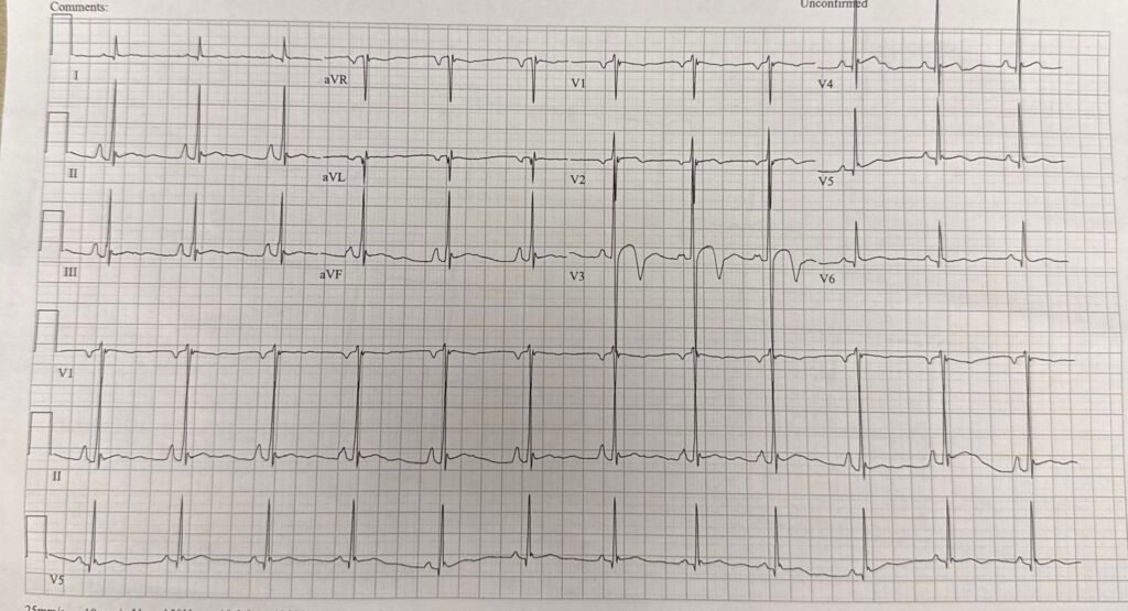
Same findings.
The first hs troponin I returned at 9 ng/L. It peaked at 41 ng/L, then fell to 29 ng/L. (I believe the URL is 34 ng/L)
Troponin need not be measured in every patient with syncope; that’s why the Canadian Syncope Rule does not require it. Syncope during exertion is a high risk feature and so measurement of troponin in this situation and admission to the hospital is quite reasonable. Moreover, sudden syncope without a prodrome is also a high risk feature (associated with a cardiac etiology), as is syncope with injury (he fell and hit his head). So, regardless of troponin, this is high risk syncope.
Acute MI of any type is defined in the Universal Definition by 1) a rise and/or fall of troponin, with one value above the 99th percentile, in the setting of ACS symptoms (or EKG findings, or imaging findings of MI). So this would fit the diagnostic criteria for acute MI (of some type) only if you believe that syncope is a symptom of ACS! Otherwise, it is classified as Non-MI “Acute Myocardial Injury”. Perhaps the low flow state of the syncope resulted in ischemia, which would be a type 2 MI but without symptoms? Type 1 MI is possible, but I think very unlikely.
OMI as a cause of Syncope: Syncope as the ONLY symptoms in OMI is quite unusual, but not unheard of. If due to OMI, syncope is almost always accompanied by some discomfort in chest, epigastrium, jaw, shoulder, arm or with dyspnea.
A repeat ECG showed no evolution:
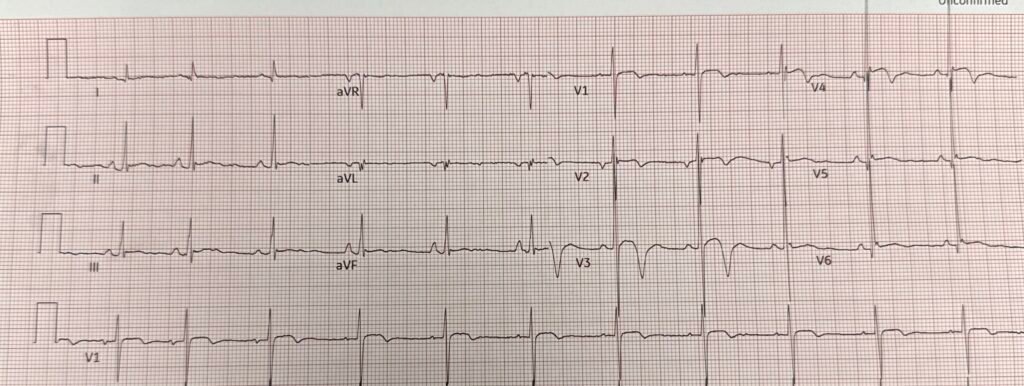
The patient was admitted and a formal echo showed:
LV: Normal with EF 60-65%, no wall motion abnormality, some increased wall thickness with “concentric remodeling.” RV was hyperdynamic.
Plan: Cardiology calls this a type 2 myocardial infarction with a differential diagnosis of hypertrophic cardiomyopathy versus other genetic abnormality. They leave hypovolemic syncope as a diagnosis of exclusion. The plan was to get an MRI later.
This post has in-depth explanation and over 20 examples of Benign T-wave Inversion (BTWI): Understanding this pathognomonic ECG would have greatly benefitted the patient.
BTWI is a normal variant associated with early repolarization. K. Wang studied it. Blinded to any patient information, Dr. Wang reviewed ECGs from all 11,424 patients who had at least one recorded during 2007 at Hennepin County Medical Center and set aside the 101 cases of benign T-wave inversion which he recognized based on the ECG alone. He then reviewed the charts: 97 were Black. 3.7% of Black men and 1% of Black women had this finding. 1 of 5099 white patients had it. Aside from an 8.8% incidence (9 of 109) black males aged 17-19, it was evenly distributed by age group.
I (Dr. Smith) reviewed these 101 ECGs, and described the key findings of the pattern:
1. There is a relatively short QT interval (QTc < 425ms)
2. The leads with T-wave inversion often have very distinct J-waves.
3. The T-wave inversion is usually in leads V3-V6 (in contrast to Wellens’ syndrome, in which they are V2-V4)
4. The T-wave inversion does not evolve and is generally stable over time (in contrast to Wellens’, which always evolves).
5. The leads with T-wave inversion (left precordial) usually have some ST elevation
6. Right precordial leads often have ST elevation typical of classic early repolarization
7. The T-wave inversion in leads V4-V6 is preceded by minimal S-waves
8. The T-wave inversion in leads V4-V6 is preceded by high R-wave amplitude
9. II, III, and aVF also frequently have T-wave inversion.
= = =
==================================
MY Comment, by KEN GRAUER, MD (7/27/2026):
I think of today’s case as, “A Case in 2 Parts”:
- Part #1 = the History ( = a 20-something man who developed syncope “while playing basketball” — but who was asymptomatic on arrival in the ED).
- Part #2: = the ECG.
= = =
The “good news” is that: i) The initial ECG (that I’ve reproduced and labeled in Figure-1) — is not the result of an acute OMI, but instead is virtually certain to represent BTWI (Benign T Wave Inversion); — and — ii) The great majority of young adults who experience syncope during exercise have a benign cause and a benign prognosis.
- The “less good news” — is that on occasion, syncope with exercise can be a “red flag” to a potentially serious (even life-threatening) underlying disorder (See below).
= = =
The Initial ECG …
As per Dr. Smith — the initial ECG in today’s case is virtually diagnostic of the normal variant pattern known as “BTWI”.
- The rhythm is sinus.
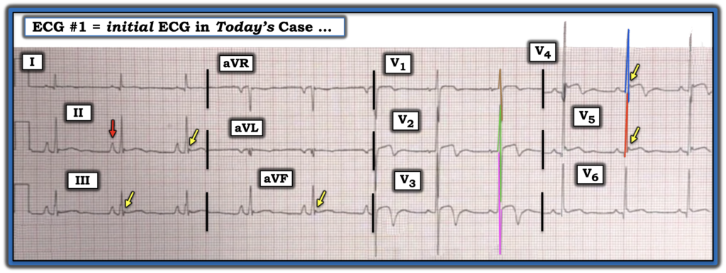
- PEARL: The tall, peaked and pointed P wave highlighted by the RED arrow in lead II of Figure-1 — represents “RAA” (Right Atrial Abnormality). But it’s important to remember that the reason the designation “RAA“ (Right Atrial Abnormality) is preferred to “RAE” (Right Atrial Enlargement) — is that tall, slender young adults (who because of their body habitus often manifest an inferior frontal plane axis) — frequently show tall, pointed P waves in the inferior leads on their ECG without there being any abnormality of the right atrium.
- In these individuals — this ECG finding of RAA is purely a result of body habitus (simply because the T wave axis often “follows” fairly closely after the QRS axis).
- Given the relatively vertical frontal plane axis in ECG #1, and the lack of other ECG signs for RVH — I suspected that the tall, peaked inferior lead P waves did not represent an anatomically abnormal right atrium. (Note the R wave in vertical lead aVF that lies at +90 degrees — is clearly taller than the R wave in horizontal lead I that lies at 0 degrees).
- Voltage is generous in ECG #1 (Note my color-coding in Figure-1 — to facilitate assessment of QRS amplitudes in those chest leads with R wave/S wave overlap).
- Regarding ST-T waves in ECG #1 — the overall “picture” is consistent with many of the 9 Criteria developed by Drs. Wang and Smith as suggestive of BTWI.
- Note #1: With experience — recognition of these criteria becomes automatic — but until that experiential point is attained, referral to the following bulleted list of these 9 Criteria may prove invaluable.
- Note #2: I’ve enclosed within parentheses after each of the criteria below — which findings are relevant to today’s ECG. (I suspect the reason for the slight variation of deepest T wave inversion that we see in today’s case [ = Criterion #3] — stems from the early precordial lead transition in ECG #1, which in turn is probably due to the concentric LVH that was seen on formal Echo).
- Note #3: For examples of 2 more tracings for which we apply these Wang/Smith criteria — Check out the June 30, 2023 post.
= = =
Applying Criteria for BTWI to Figure-1:
- Criterion #1: There is a relatively short QT interval (The QTc looks to be barely 400 msec.).
- Criterion #2: The leads with T-wave inversion often have very distinct J-waves (YELLOW arrows highlight leads with J-point notching — with deep T wave inversion in lead V4 — and subtle T wave inversion in the other J-point leads).
- Criterion #3: The T-wave inversion is usually in leads V3-V6 — which is in contrast to Wellens’ syndrome, in which T-wave inversion is usually in leads V2-V4 (Actually — The T wave inversion in today’s ECG is maximal in leads V2-V4).
- Criterion #4: The T-wave inversion does not evolve and is generally stable over time — which is in contrast to Wellens’ Syndrome, which always evolves. (T wave inversion did not evolve in today’s case).
- Criterion #5: Chest leads with T-wave inversion often have some ST elevation (ST elevation is clearly seen in leads V2,V3,V4 of today’s ECG — and these are the leads with prominent T wave inversion in today’s case).
- Criterion #6: Right chest leads often have ST elevation typical of classic early repolarization (Not present in today’s ECG …).
- Criterion #7: The T-wave inversion in leads V4-V6 is preceded by minimal S-waves (Not relevant to today’s ECG).
- Criterion #8: The T-wave inversion in lateral chest leads V4-V6 is preceded by high R-wave amplitude (Today’s tracing manifests early transition — with relatively tall R waves already present in leads V1,V2 — followed by greatly increased biphasic R/S waves in leads V2,V3 — and then tall R waves in leads V4,V5).
- Criterion #9: Leads II, III, and aVF also frequently have T-wave inversion (The inferior leads in today’s ECG manifest low amplitude ST-T waves with J-point notching, slight ST elevation — and a hint of terminal T wave inversion).
- = = =
- Criterion #10 (which I suggested in the April 23, 2026 post based on commentary by Drs. Smith and Nossen): The presence of clearly positive U Waves in an ECG from a younger adult in association with larger QRS amplitudes and “benign-looking” ST-T waves — since positive U waves in this context are generally not associated with reperfusion T waves. (I interpreted the small, rounded terminal deflections following the deep T wave inversion in leads V2,V3,V4 of today’s tracing as positive U waves).
Conclusion: As per Dr. Smith — the initial ECG in today’s case represents BTWI because: i) It “looks like” BTWI; — and, ii) Although the location of deepest T wave inversion is a bit more anterior than is typically seen with BTWI — there is plentiful voltage with no loss of chest lead R waves, with lots of benign-looking J waves marking coved ST elevation with a relatively short QTc in those leads with deep T wave inversion.
- Case Follow-up supports this conclusion:
- The demographics “fit” (The patient is a young adult African American male who had syncope but no chest pain).
- An ECG from a year earlier showed similar findings consistent with BTWI.
- Repeat ECGs showed no evolution.
- Echo showed excellent contractility without cardiomopathy.
= = =
Figure-1: I’ve labeled the initial ECG in today’s case.
= = =
Syncope with Exercise …
Today’s patient is a 20-something male who had syncope while playing basketball. He was asymptomatic by the time he arrived in the ED.
- Although many (most) cases of syncope in a younger adult who presents to the ED are benign and do not require extensive evaluation — more serious (potentially life-threatening) causes do occur.
- The most common cause of syncope is a benign vasovagal episode. This intrinsic reflex arises in response to any of a number of triggers that activate parasympathetic tone while reducing compensatory sympathic tone. The result is vasodilation without the ability to compensate for the drop in blood pressure.
- Common triggers of vasovagal syncope include emotional or physical stress (ie, extreme fear, anxiety, severe pain, exhaustion, prolonged standing, the sight of blood, and others …).
- In contrast — syncope with exercise is much less common than vasovagal syncope, but significantly more likely statistically to be associated with an adverse prognosis.
- KEY Point: We want to distinguish between: i) Syncope that occurs during exercise (which constitutes a higher risk group); — vs — ii) Syncope that occurs after exercise is stopped — which is often the result of a more benign vasovagal-mediated response (Malloy-Walton and Tisma-Dupanovic — HeartRhythm Case Rep 5(10):485-488, 2019).
- BOTTOM Line: Although today’s patient quickly recovered following his syncopal episode (As best I can tell, he was asymptomatic on arrival in the ED) — more information is needed to better determine when with respect to exercise his syncopal episode occurred (ie, during exercise, as the specific reason he fainted — or after he stopped playing basketball and was no longer active).
- As best I can tell — the patient had paused full activity (ie, He was walking over toward the side when he began to feel lightheaded; He fainted shortly thereafter when play resumedand he tried to run downcourt). This history would seem to favor a more benign vasovagal-mediated response.
- That said — more information should be obtained regarding family history of sudden death or other risk factors that might favor a poorer outcome and necessitate need for further evaluation (ie, with cardiac MRI, cath, exercise testing in an attempt to reproduce events, etc.).
= = =
= = =