= = =
Case Presentation by Willy Frick
Smith: This case reminds me of this case: 31 Year Old Male with RUQ Pain and a History of Pericarditis. Submitted by a Med Student, with Great Commentary on Bias!
Case
A 30 year old man with hypertension was brought into the hospital with chief complaint of chest pain.
EMS history: “The patient was pale, warm, and diaphoretic, sitting outside and complaining of chest pain. […] The patient was administered 324 mg of aspirin PO and 0.4 mg of sublingual nitroglycerin per protocol. Following nitroglycerin administration, the patient’s blood pressure decreased from 160/100 mmHg to 130/80 mmHg. The patient reported improvement in chest pain. The patient was transported to [hospital]. No change in the patient’s condition was noted during transport.”
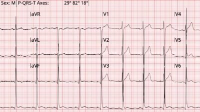
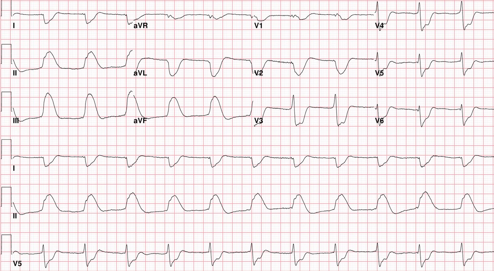
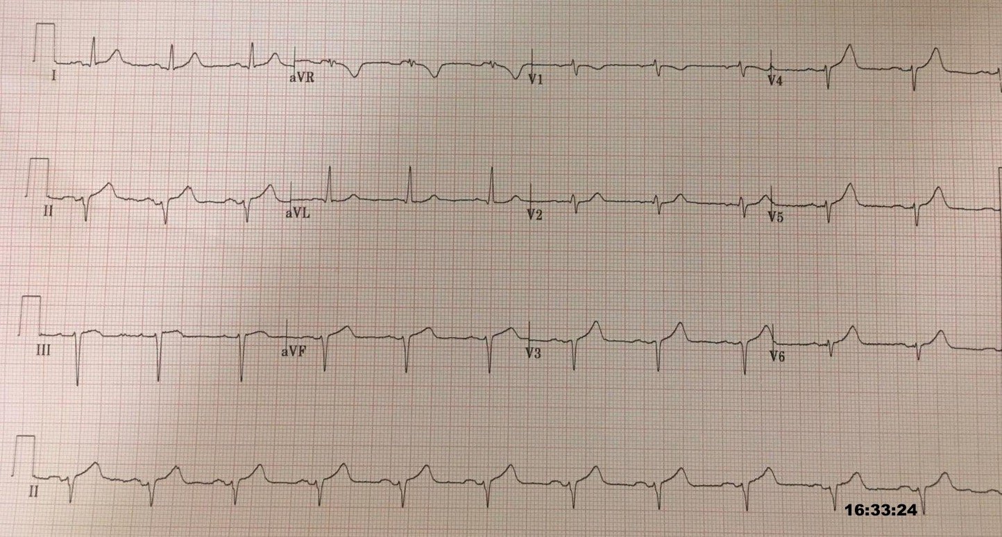
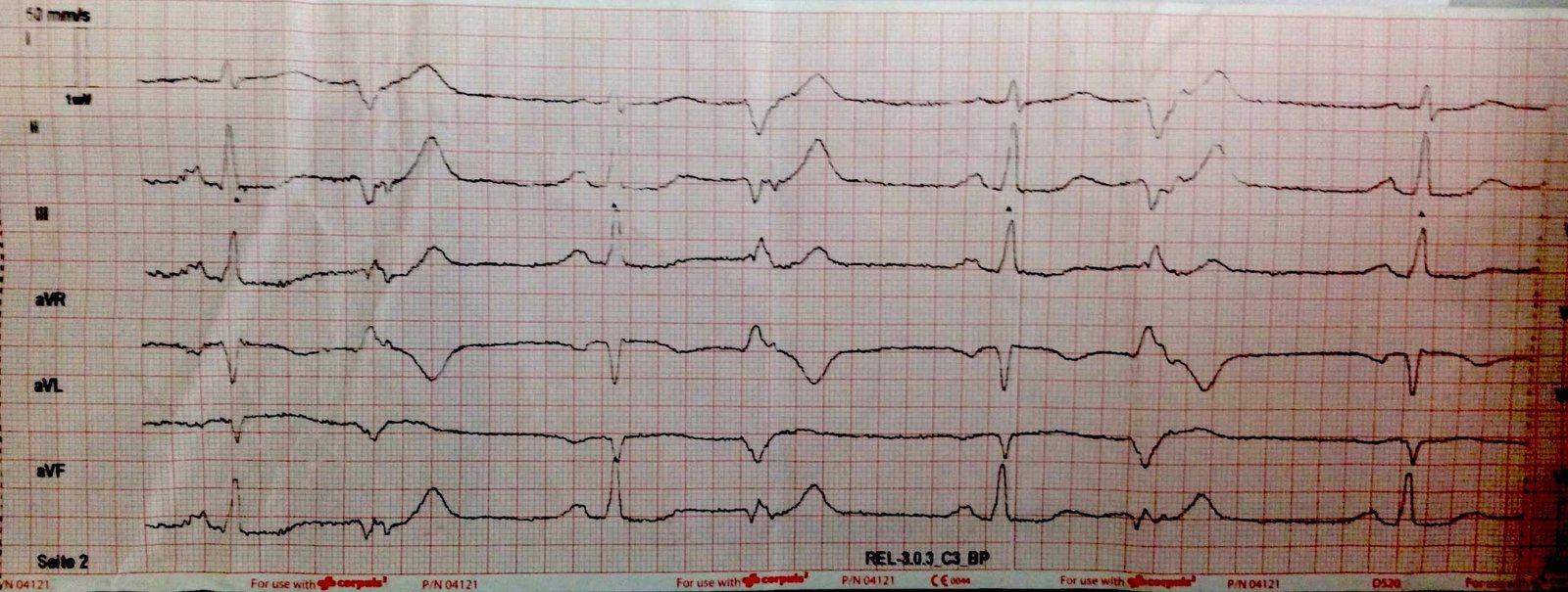
Four ECGs were obtained by the EMS crew during transport, time stamps are included for reference.
ECG 1
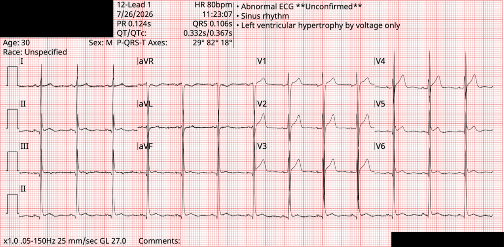
ECG 2
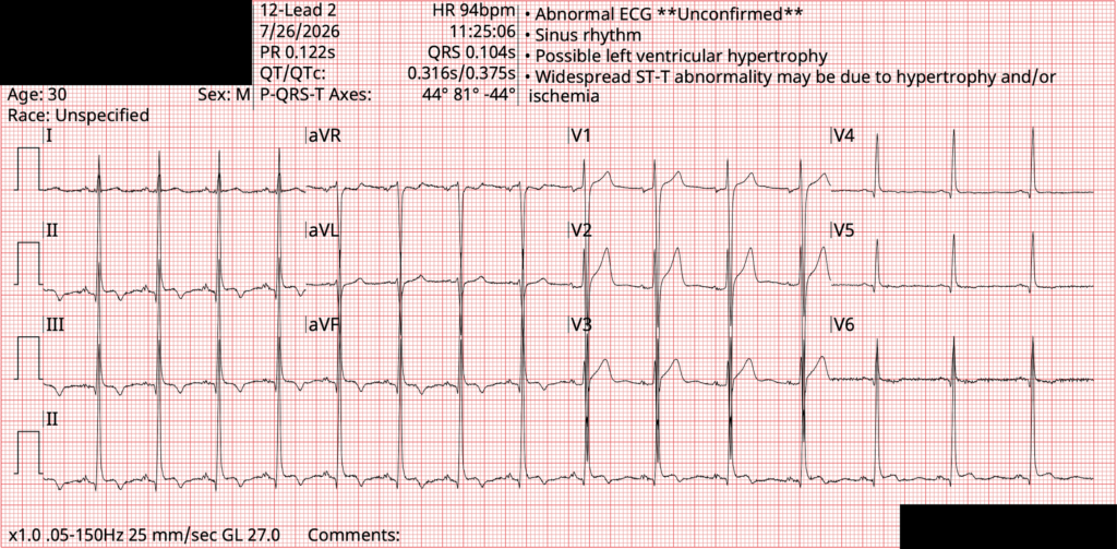
ECG 3
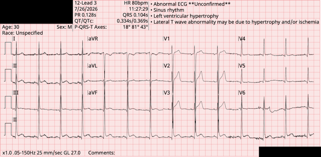
ECG 4
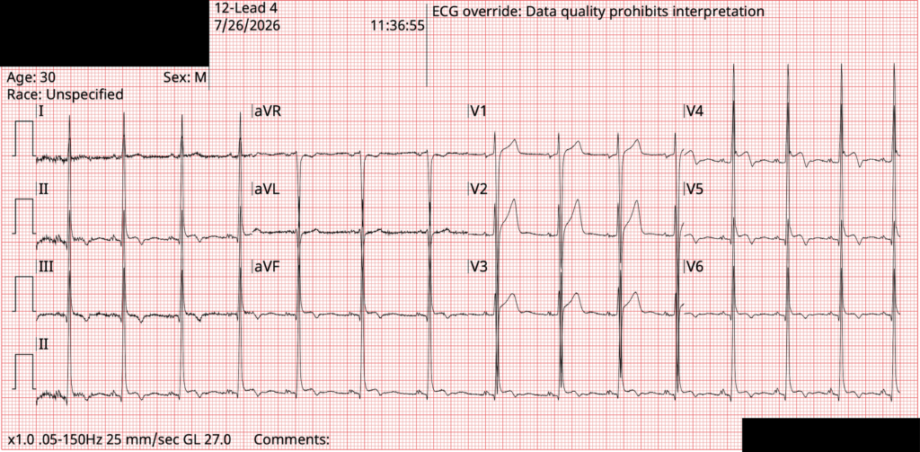
Smith: PMCardio AI ECG Model experimental rated this ECG as 76.6% early repolarization (positive) and 33.7% for pericarditis (indeterminate).
Upon arrival to the hospital, additional history is documented.
Nurse history: “Pt [brought in by EMS] c/p CP x 1 hour, given nitro and ASA by EMS. States that the pain came out of nowhere while he was at the bus stop.”
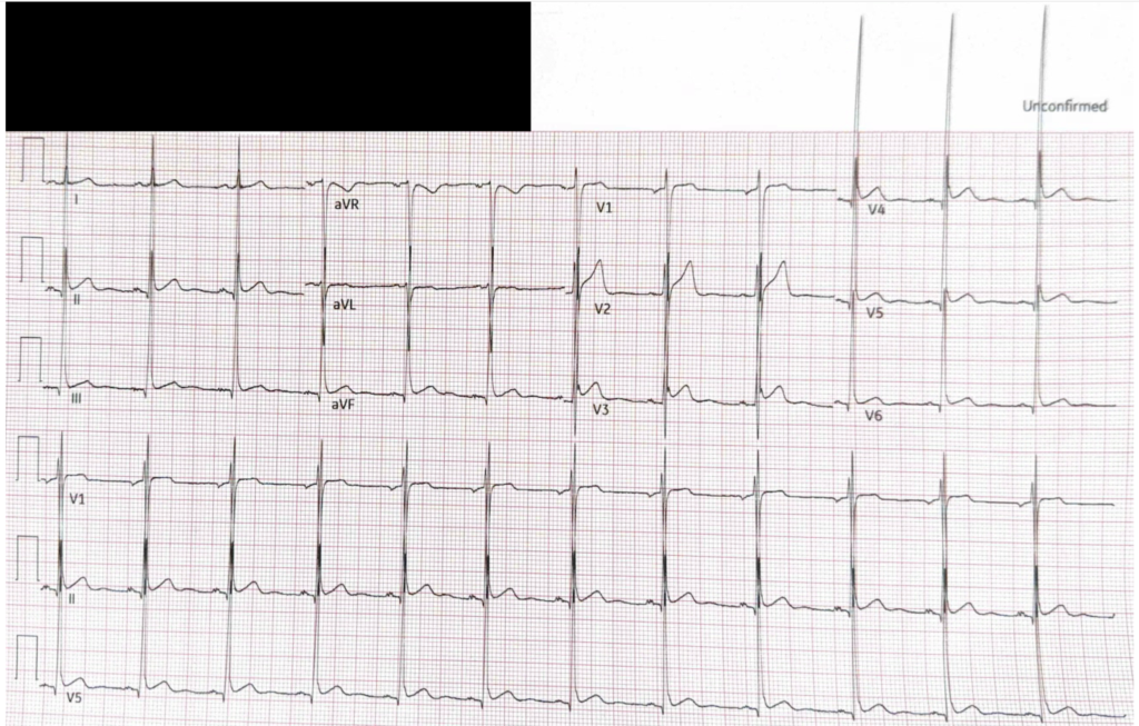
Repeat ECG was performed in the ER at 11:48, 12 minutes after ECG 4.
ECG 5
Emergency medicine physician history: “Patient states he was waiting for the bus around 1100 when he developed left-sided chest pain. Describes it as a deep pressure. Radiates to stomach. States that he’s had chest pain in the past that has come and gone, but has not felt as bad as this. Endorses diaphoresis and shortness of breath with pain.”
The emergency room physician consulted cardiology. An additional ECG was obtained at 12:37, 49 minutes after ECG 4.
ECG 6
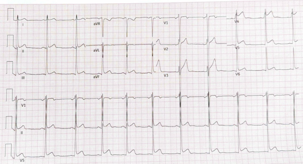
Smith: PMCardio AI ECG Model experimental rated this ECG as 88.2% early repolarization (positive) and 3.8% for pericarditis (extremely unlikely).
Cardiology history: “Patient BIBEMS for chest pain that began 3-4 hours prior to presentation. While en route with EMS he received SL NTG and ASA load with reported improvement in symptoms. In the ED, emergent cardiology consult for dynamic STE changes with STE in nearly all leads, some PR depressions, no reciprocal changes. On my evaluation he endorses 6-7/10 nonradiating left-sided chest pain that acutely worsened this morning. He states he has noticed ongoing chest pain for about the last 4 months. The pain is positional, worse with lying flat, improved with leaning forward. Pain is not worsened with exertion and he feels that it worsens with rest. He does not recall any illnesses prior to chest pain onset 4 months ago.”
The cardiology note also mentions absence of a friction rub. The patient was admitted for additional testing and observation.
Internal medicine admission history: Patient reported intermittent chest discomfort over the past several months. He noted that in the past 2 weeks he has also experienced more frequent headaches. He stated that he noticed his chest pain became more severe than in the past at about 11 AM on 7/26, ranking the pain as 10/10 in severity. He described the pain as a constant pressure that did not radiate and was worse when resting or lying flat and better when leaning forward. He also endorsed fever/chills and shortness of breath on the day of admission in association with his chest pain.”
Before reading further, what do you think is the correct diagnosis?
A. Acute myocardial infarction
B. Pericarditis
C. Myocarditis
D. Other
Case continues…
The cardiology consultant suspected pericarditis and recommended treatment with aspirin and colchicine along with transthoracic echocardiogram and inflammatory markers (ESR/CRP).
- Five serial high-sensitivity troponin I measurements were undetectable
- NT-proBNP was undetectable
- ESR was 6 (ref: 1-15)
- CRP was 0.7 (ref: 0.7)
- Echocardiogram showed normal LVEF and normal wall motion, and “normal pericardium without pericardial effusion”
Finally, here are the patient’s previously recorded ECGs spanning the prior 9 years. Keep in mind that “Benign T-wave Inversion” is a form of Early Repolarization.
2017
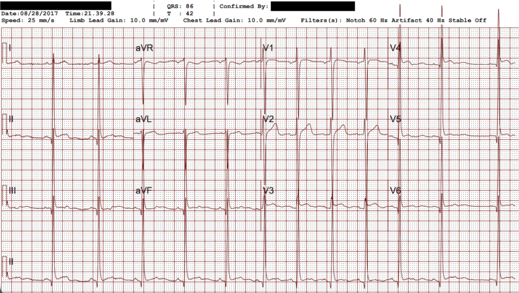
2019
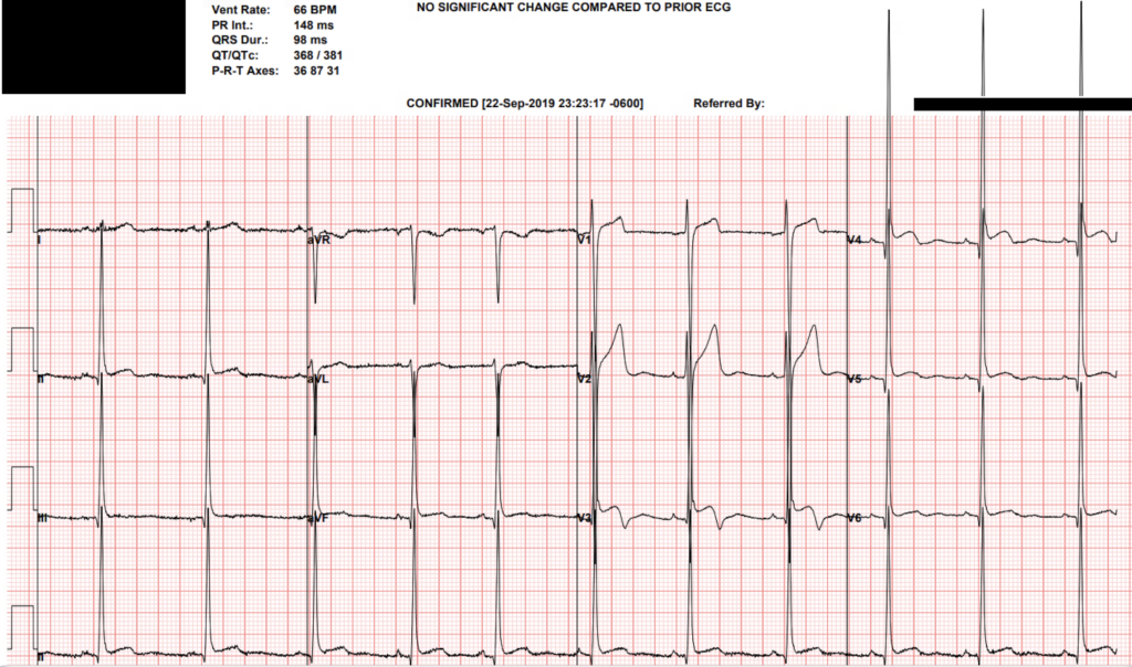
Smith: PMCardio AI ECG Model experimental rated this ECG as 78.8% early repolarization (positive) and 24.5% for pericarditis (negative).
2020
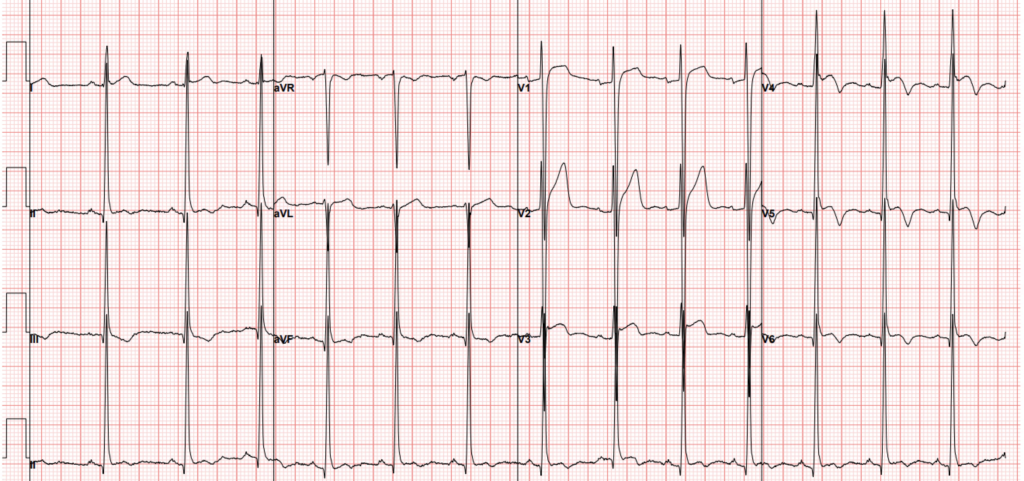
Smith: PMCardio AI ECG Model experimental rated this ECG as 65.2% early repolarization (positive) and 21.9% for pericarditis (negative).
2022
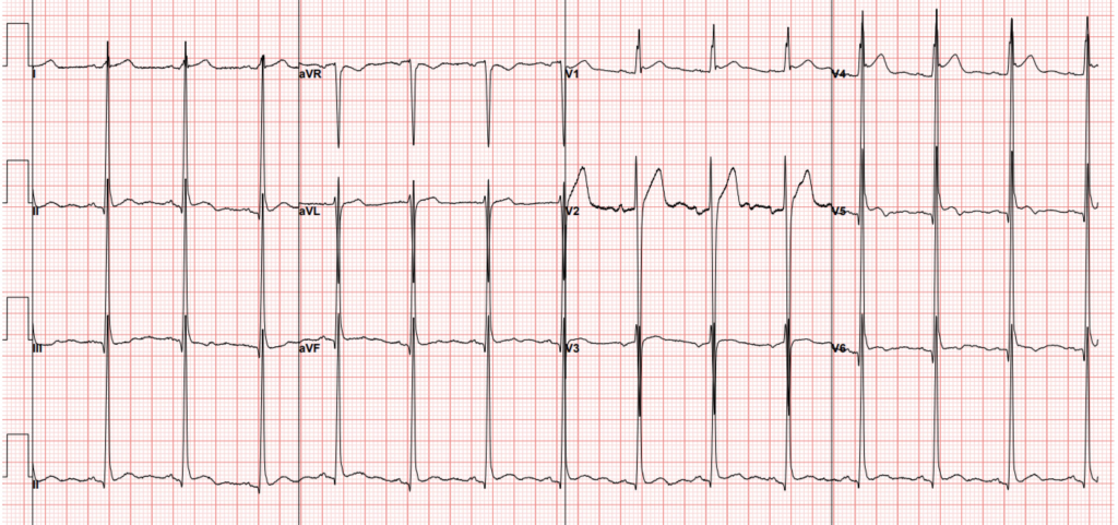
2023
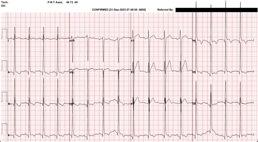
The final diagnosis reached by the treating clinicians was pericarditis, and the patient was discharged with 3 months of colchicine and a short course of ibuprofen.
Returning to the question posed above, what is the correct diagnosis?
——-D. Other. This is a normal variant ECG.
The cause of the chest pain is not clear, but it has nothing to do with the heart or ECG.
Discussion
The blog has published innumerable cases of this normal variant known as “benign T wave inversion.” Here is a recent case, and here is a more extensive catalogue of similar cases.
We have reviewed this in prior posts, but it is worth saying in clear, explicit terms:
- This ECG is NOT ischemic
- This ECG does NOT represent pericarditis
- This ECG does NOT represent LVH
It is a frustrating and confusing FACT that a patient can present with dynamic ECG changes and ST elevation, but still not have any acute cardiac pathology.
The STEMI paradigm has poisoned our brains into thinking that all ST elevation is pathological, and this corrosive idea is at the crux of the diagnostic error in this case.
I think there are at least two additional good lessons here in patient care.
1. It is easy to elicit the history that confirms your suspicions.
We see a clear evolution in documented history here.
- The EMS and nursing histories indicate acute chest pain with diaphoresis, improving with nitroglycerin, consistent with a perceived diagnosis of ACS.
- The emergency medicine history describes left-sided chest pain radiating into the stomach with diaphoresis and dyspnea. The pain is described as a deep pressure, once again consistent with ACS.
- The cardiology history has changed significantly, now more acute on chronic without radiation, and the pain is worse with supination. Much more consistent with pericarditis.
- The internal medicine history has some features of both — acute on chronic pressure-like pain worse with rest and supination, and with associated fevers and chills.
The point is NOT that some clinicians took good histories and others took bad histories. But that history taking is a two-party process (clinician and patient) with bidirectional influence. It is an inherent human bias to ask leading questions. We were not present when the above clinicians took history, so we do not know if they asked leading questions or not.
- A leading question is: “Does the pain get worse when you lie down?”
- A non-leading question is: “Does anything make the pain worse?”
It is worth remembering that history is a diagnostic test, and like all diagnostic tests it is imperfect.
2. Anchoring bias is a powerful source of diagnostic error.
In this case, the clinicians treating the patient may not have been blog readers, or if they were they forgot about benign T wave inversion. ECG is hard. Most clinicians will not make this diagnosis by ECG, and that’s a fact. With the emergence of AI, our ability to make diagnosis by ECG will improve significantly. (QOH called not OMI on all ECGs). Surely within a few years we will have models that can diagnose or reject pericarditis with good accuracy.
But BOTH pericarditis and OMI are clinical diagnoses which are not defined by ECG. In this patient’s case we had many reasons to reject the diagnosis of pericarditis.
- Normal inflammatory markers
- Absence of pericardial effusion
- Absence of friction rub
- Pressure-like pain (pericarditis should be sharp)
- Stability (or at least meta-stability) of the ECG over 9 years
AI will not be the solution to cognitive bias, in fact it may reinforce our biases, especially since it requires large volumes of annotated data for training. If cardiologists are calling this ECG consistent with pericarditis, I worry what that could mean for future AI models.
Learning points
- Learn to recognize this normal variant ST elevation ECG.
- Keep history questions open ended, try not to lead the patient.
- As you accumulate data, return to your differential diagnosis and ask yourself what fits and what does not.
- Early repolarization may be dynamic! Not all evolution and not all increasing ST Elevation is ischemic evolution or STE. See these blog posts/cases with two supporting references:
= = =
==================================
MY Comment, by KEN GRAUER, MD (7/30/2026):
Important case by Dr. Frick! Many points are highlighted:
- If you are looking for a certain diagnosis — You are more likely to elicit findings that support your searched-for diagnosis.
- Acute pericarditis is not common. When the prevalence of a certain disease is low — then a majority of equivocal cases will be false positives. In contrast, when the prevalence of a disease is much higher — then a signficantly greater percentage of equivocal cases will turn out to be true positives.
- Although I found myself looking hard for ECG abnormalities — I did not think the multiple serial ECGs in today’s case were diagnostic of either acute pericarditis or acute OMI. (See discussion of Figure-1 below).
- Instead, what impressed me most about the multiple tracings recorded in today’s case — is the variability in heart rate and in pecordial lead placement. For example — T wave inversion especially increases in the inferior leads in ECG #2, in which the heart rate is at its fastest. The effect of variable precordial lead placement is perhaps best appreciated by scrolling through the 6 serial ECGs with focus on lead V4, which shows an R wave amplitude as small as 5 mm (in ECG #6) — but as great as 40 mm in ECG #5. Bottom Line: I did not think the modest differences in QRST morphology seen over the course of the 6 serial tracings in today’s case qualified as true “dynamic” changes.
- NOTE: There is a definitive test for acute pericarditis = Cardiac MRI (especially with LGE = Late Gadolinium Enhancement). Given uncertainty about the diagnosis in this 30-year old man who has had multiple ECGs performed over the previous 9 years — assessment by cardiac MRI would seem indicated (Nice review by Baser et al on the use and diagnostic value of cardiac MRI for identifying acute pericarditis at this link — Methodist Debakey Cardiovascular J: 22(2):41-49, 2026).
= = =
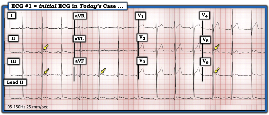
Today’s initial ECG …
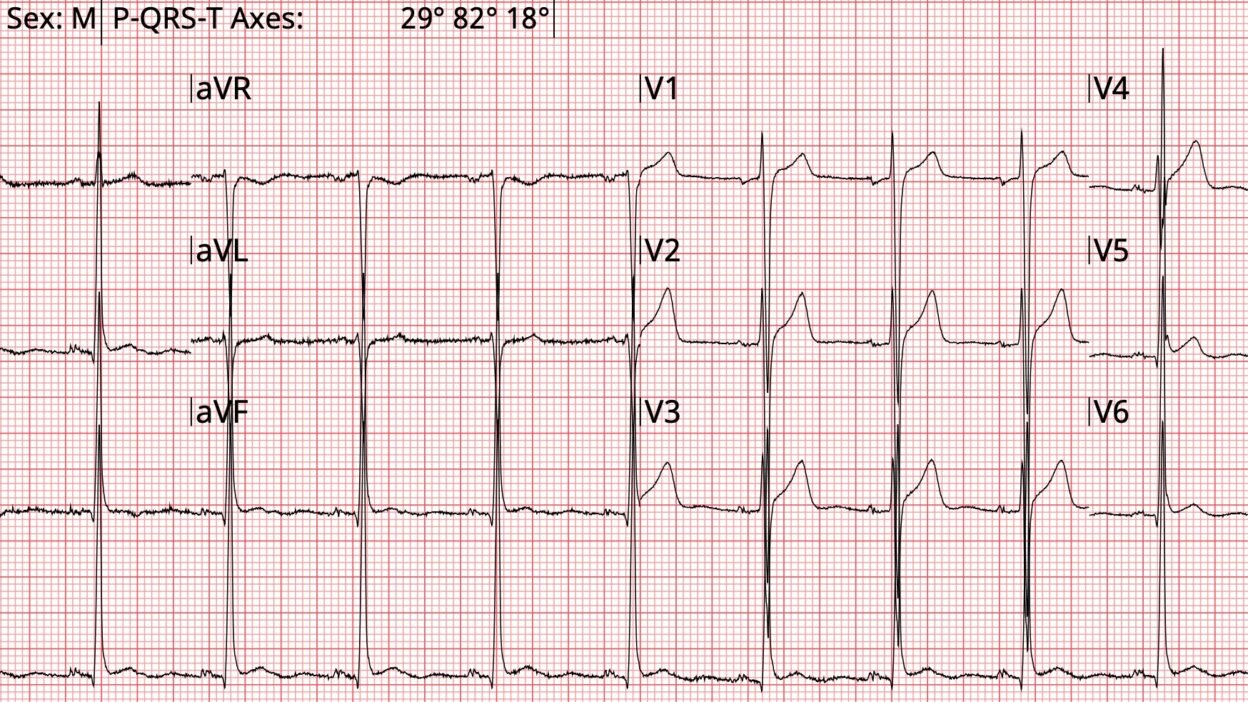
Assessment of this patient’s serial ECGs is complicated by his history of hypertension that predisposes him to LVH. That said — there are features suggestive of a repolarization variant on this patient’s serial tracings. As I allude to above — these features vary somewhat from one tracing to the next (due at least in part to a variable heart rate and inconsistent precordial lead placement). For simplicity — I limit my comments to the initial ECG (recorded at 11:23:07 in today’s case) — which I’ve reproduced in Figure-1. I felt the subsequent 5 tracings reflect variations on the “theme” that we see in ECG #1. Note the following in this initial ECG:
- QRS amplitude is markedly increased, with overlap of R waves with S waves in multiple leads. As a result of these large QRS amplitudes — the ST elevation that is seen does not appear excessive.
- R wave progression is normal.
- The QTc looks to be relatively short.
- Slurring of the terminal portion of the QRS (with “slurring” generally accepted as an alternative to J-point notching as a repolarization variant sign) — is seen seen in multple leads (YELLOW arrows in Figure-1).
Additional thoughts on today’s case:
- T wave inversion is an intermittent feature in today’s 6 serial tracings — but when present, it is associated with markedly increased QRS amplitudes, terminal QRS “slurring”, and ST elevation in the same leads that manifest T wave inversion. This composite picture is best appreciated in lateral chest leads V4,V5,V6 of ECG #4 — that manifest an appearance that “looks” benign.
- For more on the complexity of early repolarization patterns and the subtlety associated with terminal QRS “slurring” — Check out My Comment at the bottom of the page in the February 5, 2026 post.
- In contrast, with acute pericarditis — T wave inversion typically is not seen at a point in time in which there is still ST elevation. So the prominent T wave inversion seen in ECGs #2 and 4 in association with ST elevation in these same leads is another point that argues against acute pericarditis as the diagnosis.
= = =
Figure-1: The first of many ECGs done in today’s case.
= = =
Additional “Take–Home“ Concepts:
- Repolarization variant tracings sometimes show changes in ST-T wave morphology over the course of serial tracings. These are not ischemic dynamic changes.
- It is essential to ensure that the frontal plane axis and chest lead appearance (ie, R wave progression & relative QRS amplitudes) are similar when comparing serial ECGs. Otherwise you are not comparing “apples with apples” — but instead are mixing in “oranges”. In my experience — this concept is all-too-often overlooked (even by experienced interpreters) when comparing serial tracings.
- The ECG picture of acute pericarditis evolves over time — but not over a period of a few minutes (as is suggested by comments of clinicians in today’s case). Variation in precordial lead placement seems a much more likely explanation for the surprisingly marked differences in chest lead appearance between some of the serial tracings.
- In addition to simultaneous T wave inversion and ST elevation — other ECG findings appear on today’s 6 serial tracings that are not typical for acute pericarditis. These include: i) The terminal QRS “slurring” in multiple leads that is so commonly seen with repolarization variants; — ii) A closer resemblance in ST-T wave morphology of lead II with lead III in some of the serial tracings (instead of with lead I); — and, iii) The reduced ST/T wave ratio (See My Comment at the bottom of the page in the June 15, 2025 post — for more on “My Take” re ECG findings in Acute Pericarditis).
= = =
Final Thoughts …
So — WHY does today’s patient continue to have such severe (up to 10/10) chest pain?
- This CP has seemingly been persistent over time, in view of multiple repeat ECGs in his chart recorded over the past 9 years. I suspect that this patient will continue to present to EDs for his symptoms unless (until) we can determine the cause of his at times very severe CP.
- Realizing that we do not know more details in this case — I’ll speculate that the cause of this patient’s CP is most probably either: i) Musculoskeletal; — or ii) More likely, psycho-social — with chronic anxiety, panic, depression, or substance abuse as the likely “culprit”.
If I was seeing this patient in my office — I would:
- Do a simple exercise treadmill test in my presence — hoping to encourage this patient to as close as possible attaining his maximal predicted heart rate on the treadmill (Looking see if this results in any abnormal ST-T wave changes with the same precordial lead placement — as well as hoping to see if I could reproduce his symptoms during some level of exercise);
- I’d take a complete psychosocial history — looking for elements of chronic anxiety, panic, depression, and/or substance abuse.
= = =