Written by Matthew McArthur
I saw a 40 year old female who had a ~3 minute, witnessed loss of consciousness at home a little after midnight. Her partner states she suddenly lost consciousness, her eyes rolled back, she convulsed several times, frothed at the mouth, became very pale, and then developed agonal respirations and stopped breathing. He called EMS. The entire episode lasted about 3 minutes and she regained consciousness prior to EMS arrival. She was awake and GCS 15 on EMS arrival with normal vitals. She is a fentanyl user who endorsed smoking fentanyl earlier that evening, and who was on high doses of prescription methadone and hydromorphone for Opioid Use Disorder. She had been on these medications for a couple years with no recent medication changes. Otherwise well with no acute illness, nausea, vomiting or diarrhea.
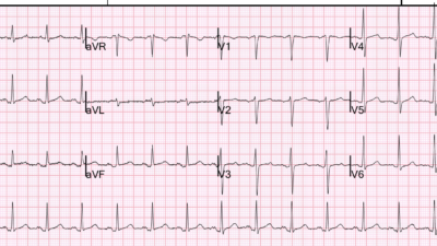
Have a look at her initial ECG:
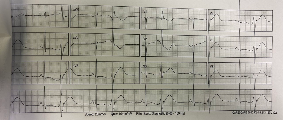
Her initial ECG is clearly abnormal: sinus rhythm with PVCs/bigeminy and long QT. Look closely at the T waves after the sinus beats, which are abruptly interrupted by the PVC. This ECG demonstrates R on T phenomenon and is high risk for imminent Torsades de Pointe.
On examining this ECG and with that story I empirically gave IV magnesium sulfate, and had her placed on telemetry and had defibrillator with pads placed on her.
Smith: The PVCs are quite bizarre with huge inflated T-waves. See the link below for even more bizarre T-waves which are associated with this pathology.
Prior visits
Further history revealed that she had been having <1 minute sudden syncopal episodes, sometimes with urinary incontinence, tonic posturing, and myoclonic convulsions (partner concerned about seizures), on and off for the last 2 weeks, about 10 episodes in total. Outside of the episodes she was asymptomatic and felt at her baseline. She was evaluated at a local ED 10 days prior, after one of these episodes — her partner brought her in concerned she had had been having seizures. On this visit she was discharged home with unremarkable bloodwork and CT head, with the ED Physician indicating he suspected these were episodes of altered level of consciousness secondary to illicit drug use as the patient endorsed recreational use of fentanyl.
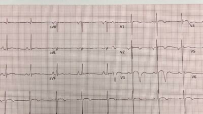
Here was the ECG at the time.
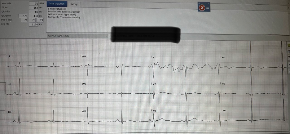
The computer reads the QTc as 438, but it looks to me that the T waves are low amplitude and biphasic (especially obvious in V4-V5), which is messing up the computer read, and the QTc is clearly MUCH longer than 438; accounting for the biphasic T wave we can see the QT interval is well over 3 big boxes (600ms). This was not noted by the ED Physician 10 days prior.
Smith: Morever, when the heart rate is below 60, QT correction SHORTENS the QTc. It is not recommended to correct for heart rate below 60. The QT nomogram, which is specifically for acquired QT due to drugs, does not correct for slow heart rate. Moreover, when the patient has a slow heart rate, they are more susceptible to “pause dependent” Torsades de Pointes. See this post for everything you want to know about the QT interval: QT Correction Formulas Compared to The Rule of Thumb (“Half the RR”)
Also:
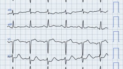
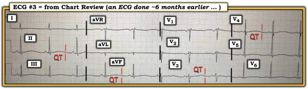
Chart review revealed that she was seen at a local ED 6 months prior for syncopal episodes. Again discharged with the ED Physician suspecting the loss of consciousness were related to illicit drug use. On this visit, her ECG showed the following.
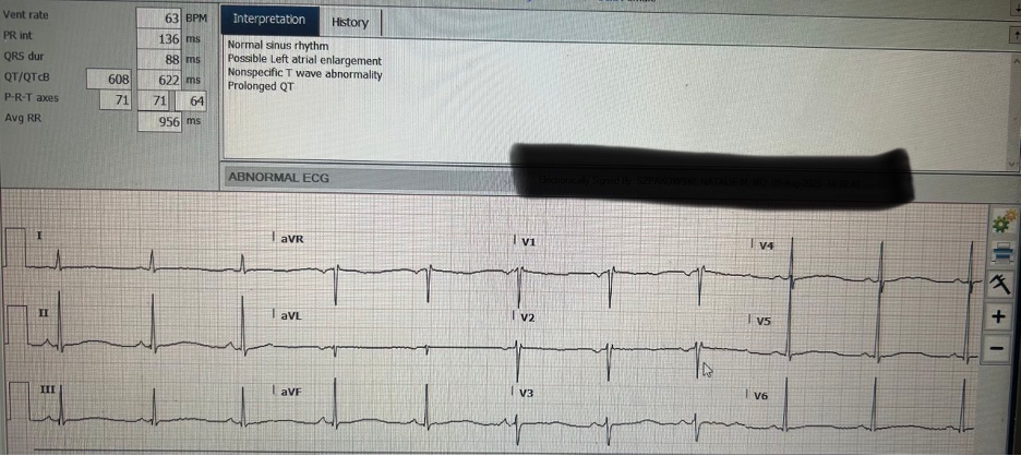
The long QT, which was this time correctly identified by the computer, was not noted by the ED Physician at the time.
Smith: Much of the time, the computer does not accurately measure the QT interval and gives false negatives. Always measure the QT yourself!!
See these cases which show how badly the conventional algorithm measures the QT and what the consequences are:
A New Seizure in a Healthy 20-something
Syncope in a 20-something woman
Syncope and Bradycardia
Back to the case
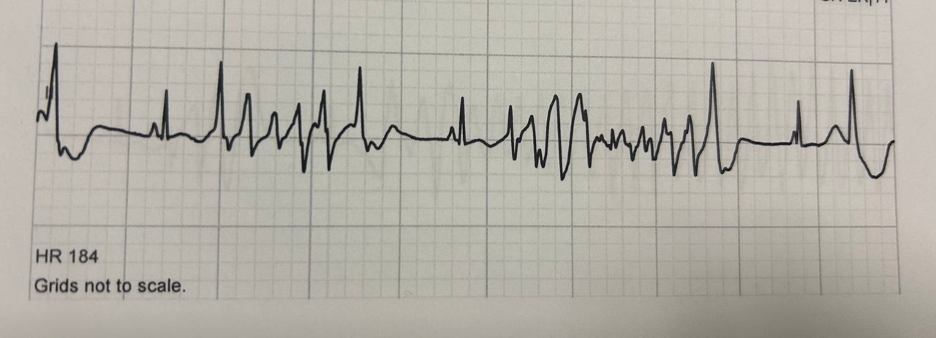
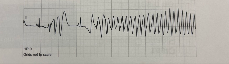
She had a transient <20s syncopal episode in the ED. Telemetry confirmed Torsades de Pointe which resolved spontaneously without any defibrillation.
Here is her telemetry strip around the syncopal event:
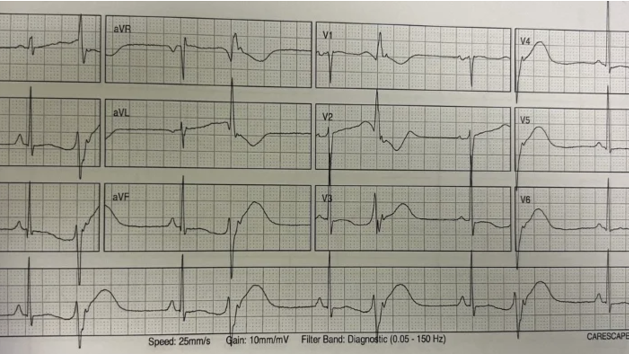
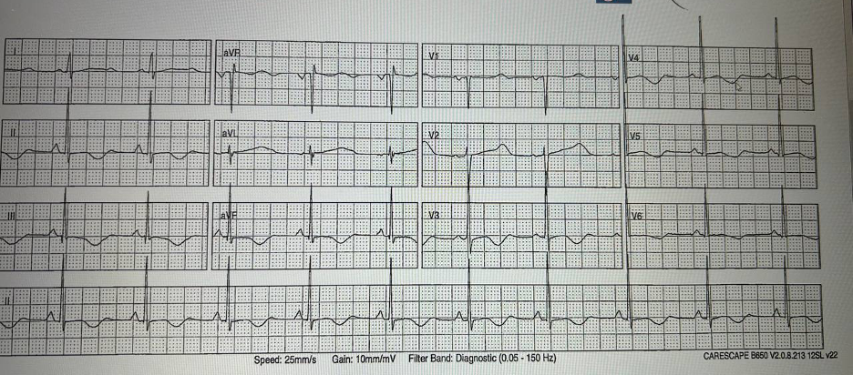
Here is an ECG after her run of torsades without PVCs showing her long QT again, with biphasic T waves seen much more clearly.
Magnesium was within normal limits and potassium slightly low (3.1), she received doses of both Mg and KCl under my care.
I consulted the nearby cardiac centre for transport to the Cardiac Care Unit (10 minute drive). Patient had a TdP arrest shortly after arrival to the CCU that was successfully defibrillated with one shock. Transvenous pacer was inserted for overdrive pacing.
The patient was weaned off methadone and the QT normalized over several days, at which point the transvenous pacer was removed and the patient was discharged.
To acutely prevent torsades recurrence, if I were managing the case again I would have started overdrive pacing with isoproterenol infusion in the ED awaiting cardiology admission, as described in this recent post .
Take-home points
- scrutinize ECG in unexplained LOC: if long QT given empiric magnesium and prepare for overdrive pacing
- methadone as cause of long QT
- computer can mess up QTc
- don’t dismiss LOC as due to substance use, get a good history
- syncope vs seizure (even in obvious cases of syncope, lay witnesses are often concerned about seizure due to witnessing myoclonic jerks, incontinence, eyes rolling back, etc). See notes below:
A note on tonic posturing and myocolonic Jerks
I have already lost count of how many times in my young career, patients’ families have witnessed a syncopal event, including multiple V Fib arrests, and told me they are worried their loved one had a seizure, mostly due to the tonic posturing and myoclonic jerks that are extremely common in syncope.
This study in Neurology published in 2018 compared video and EEG recordings of 65 cases of tilt-table induced syncope and 50 cases of seizure at an academic centre in the Netherlands. Tonic postures were present in 65% of syncopal episodes and myoclonic jerks were present in 51% of syncopal episodes. In their study, no seizure had < 10 myoclonic jerks, and no syncopal event had > 20 leading the authors to propose the 20/10 rule to differentiate seizure vs syncope based on the number of myoclonic jerks witnessed.
After the myoclonic jerks and tonic posturing, all syncopal episodes progressed to atonia, whereas none of the seizures had atonia. The myoclonic jerks in seizures were more rhythmic than those in syncope.
A note on urinary incontinence in syncope
It is often taught that urinary incontinence increases the likelihood of seizure, however urinary incontinence can happen in a variety of different causes of loss of consciousness. This systematic review and meta-analysis in Seizure in 2013 looked at studies evaluating the presence of urinary incontinence in both epileptic seizures and non-epileptic mimics (syncope or psychogenic non epileptic seizures). This study found that the presence of urinary incontinence has no diagnostic value for differentiating seizure from syncope or non epileptic seizure.
Another case of long QT from Methadone: What are these bizarre bigeminal PVCs??
Look at all these Bizarre PVCs (many examples at this link): What are these bizarre bigeminal PVCs??
= = =
======================================
MY Comment, by KEN GRAUER, MD (7/21/2026):
I found the ECGs in today’s case to be challenging. That said — I arrived at a similar conclusion to that described by Drs. McArthur and McLaren, albeit with slight difference in my path for getting there.
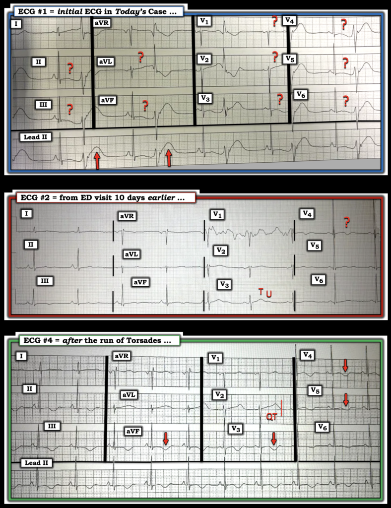
- In the interest of academic discussion — I focus my comment on my different path. I illustrate my perspective in Figure-1 — in which I’ve reproduced 3 of today’s ECGs (with a retrospective look at the 4th 12-lead ECG that was done on this patient in Figure-2).
= = =
What is the QTc in Today’s Tracings?
I found it especially difficult to determine the “true” QTc in 3 of today’s 4 12-lead tracings. That said — how long the corrected QT interval truly is in these 3 tracings does not really matter — because:
- i) Most opioids can prolong the QTc — especially when these drugs are taken in large quantities (Hu et al — Cardiovasc Toxicol 24(5):472-480, 2024) — and — consumption by today’s patient of known “high doses” of prescription methadone and hydromorphone (for Opioid Use Disorder) — in association with this patient’s admission of recreational fentanyl on the evening that ECG #1 was recorded magnifies the potential for QTc prolongation and associated cardiotoxicity;
- ii) Associated electrolyte imbalance is synergistic to the QTc prolongation effect of 3 high-dose opioids (and synergistic in increasing cardiotoxicity) — with documentation of significant hypokalemia ( = 3.1 mEq/L) on the day ECGs #1 and #3 were recorded — and with U waves suggesting low K+ on the day ECG #2 was recorded.
- iii) Drs. McArthur and McLaren very nicely documented the rhythm strip during an episode of Torsades de Pointes (with strong suggestion by history of numerous syncopal episodes — some number of which seem likely to represent more Torsades).
- iv) Given documented Torsades (with suspicion of multiple recurrences of this arrhythmia) — bradycardia on ECG — and ingestion of high doses of 3 opioids known to prolong the QTc, potentiated by hypokalemia ==> the indication for K+ replacement and IV Mg++ as Torsades treatment measures is secure regardless of what the QTc duration happens to be.
- v) Any marked QT prolongation that might be present will probably go away following K+ replacement, IV Mg++ treatment, and prudent reduction of opioid dosing as is possible.
= = =
The 3 ECGs in Figure-1:
- ECG #1: The rhythm is ventricular bigeminy (Note 1:1 retrograde VA conduction marked by the negative notch after each PVC in the long lead II). I do not see clear delineation of the end of the T wave for sinus beats in any of the 12 leads (RED question marks). Instead — ST-T waves are nearly amorphous (flattened). Therefore — I did not know how to measure the QT interval.
- The above said — I strongly suspected QT prolongation because of the predisposing opioids and the hypokalemia.
- Speculation by ME: I thought the size and shape of the T waves of the PVCs in ECG #1 was fascinating! Over the years, we have published a number of cases of acute OMI in which ST-T waves were nonspecific for sinus-conducted beats — but diagnostic of acute OMI from the ST-T waves of PVCs! Although I’ve not seen this reported — I suspect a similar phenomenon occurring here, in that the T waves of each PVC in ECG #1 look disproportionately huge, with what has to be a markedly prolonged QTc that simply was not evident from assessment of sinus-conducted beats.
= = =
Figure-1: To facilitate comparison — I’ve labeled and placed 3 of today’s 4 12-lead ECGs together.
= = =
- ECG #2: The rhythm is sinus bradycardia at ~50/minute. Artifact mars lead V1. With the exception of leads V3 and V4 — there is little shape to most ST-T waves. This once again makes determination of the end of the T wave in most leads difficult. That said — I thought lead V3 remarkable for the presence of a T wave and a U wave — and considering the bradycardia, I didn’t think the QT interval was necessarily abnormal (ie, There was “Q-U” prolongation — but to my eye, not ncessarily QT prolongation). I was less sure how to interpret the negative deflection in lead V4 given artifact elsewhere and no other chest leads with this negative deflection.
- NOTE: We have added a QTc Calculator to the Top Menu of every page in Dr. Smith’s ECG Blog (— CLICK HERE — found under the Tools & Guides tab). As becomes obvious from the 5 most used formulas for QTc calculation that we show — universal agreement is lacking, and there is no perfect method for QTc calculation. Unfortunately — the accuracy of QTc estimation is especially reduced with slow heart rates — which clinically becomes less important because bradycardia itself predisposes to Torsades. That said — I took lead V3 in ECG #2 as providing my most reliable QT duration landmarks, and I therefore did not estimate the QTc as more than borderline given the slow heart rate of ~50/minute.
- = = =
- ECG #4: There is sinus rhythm at ~60/minute. We finally see clear delineation of ST segments and T waves in this tracing — and the QTc is markedly prolonged. That said — this patient just had a run of Torsades prior to the recording of this ECG. How much of these hypervoluminous T waves in ECG #4 might be the result of a “Memory” effect from this brief episode of Torsades — rather than baseline QTc prolongation? (especially since ST-T waves in the previous ECG done a little bit earlier in this patient were flattened — as is seen in ECG #1).
= = =
Another Look at the 3rd 12-Lead ECG …
Knowing “the Answer” (ie, Torsades de Pointes) — I went back and took another look at the 3rd 12-lead ECG that was shown in the above discussion by Drs. McArthur and McLaren in today’s case (ie, this being the ECG done 6 months earlier at a local ED).
- As seen in Figure-2 — ST-T waves in this tracing (that was done 6 months earlier) are once again all-but-amorphous from ST-T wave flattening — BUT — there does appear to be a definable end to the T wave (perhaps seen best in lead V3). Given the sinus rhythm heart rate of ~60/minute — the QTc will be the same as the QT interval that we measure, which looks to be nearly 600 msec.
- = = =
- Additional “Take-Home” from Today’s CASE:
- i) Assessing the QTc is sometimes not a simple task.
- ii) Looking through the chart at prior tracings can be enlightening.
- iii) Precipitants of Torsades are often multifactorial ( = 3 opioids, hypokalemia, and bradycardia in today’s case).
- iv) Normalizing K+ and IV Mg++ are key treatments.
= = =
Figure-2: The ECG from 6 months earlier.
= = =
= = =